Computer system vision, which allows computer systems to pull information from digital images, is a type of maker knowing that has actually been around for decades, so he and his coworker and fellow corresponding author Dr. Peter Bubenik, a mathematician at the University of Florida and an expert on topological data analysis, chose to partner the information of microscopy with the science of topology and the analytical might of AI. “We want to bring computer objectivity to it and we want to bring a higher degree of pattern acknowledgment into the analysis of images.”
Some of those device knowing designs that require hundreds of images to train and classify images dont describe which part of the image contributed to the classification, the detectives write. The new system instead needs comparatively few high-resolution images and defines the “patches” that led to the picked category. In a handful of minutes, the scientists basic individual computer can complete the new image analysis pipeline.
” We believe this is interesting development into using computer systems to provide us new information about how image sets are various from each other,” Vitriol states. “What are the real biological modifications that are happening, consisting of ones that I might not be able to see, due to the fact that they are too minute, or since I have some kind of predisposition about where I ought to be looking.”
At least in the analyzing information department, computers have our brains beat, the neuroscientist says, not just in their objectivity but in the quantity of information they can examine. Computer vision, which enables computers to pull information from digital images, is a type of artificial intelligence that has been around for decades, so he and his colleague and fellow corresponding author Dr. Peter Bubenik, a mathematician at the University of Florida and a professional on topological data analysis, decided to partner the detail of microscopy with the science of geography and the analytical may of AI. Topology and Bubenik were key, Vitriol states.
Geography is “best” for image analysis since images include patterns, of items arranged in space, he states, and topological data analysis (the TDA in TDAExplore) assists the computer system likewise recognize topography, in this case where actin– a protein and essential building block of the fibers, or filaments, that aid provide cells shape and motion– has moved or changed density. Its an efficient system, that instead of taking literally numerous images to train the computer how to recognize and categorize them, it can learn on 20 to 25 images.
Part of the magic is the computer system is now discovering the images in pieces they call spots. Breaking microscopy images down into these pieces makes it possible for more precise category, less training of the computer on what “typical” looks like, and ultimately the extraction of significant data, they write.
No doubt microscopy, which makes it possible for close examination of things not noticeable to the human eye, produces lovely, comprehensive images and dynamic video that are a mainstay for numerous scientists. “You cant have a college of medication without advanced microscopy centers,” he states.
But to first understand what is typical and what happens in illness states, Vitriol requires comprehensive analysis of the images, like the number of filaments; where the filaments remain in the cells– near the edge, the center, scattered throughout– and whether some cell areas have more.
The patterns that emerge in this case tell him where actin is and how its organized– a significant element in its function– and where, how and if it has altered with illness or damage.
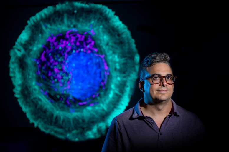
As he takes a look at the clustering of actin around the edges of a main nerve system cell, for example, the assemblage informs him the cell is expanding, moving about and sending projections that become its leading edge. In this case, the cell, which has been basically dormant in a meal, can spread out and stretch its legs.
Some of the problem with researchers examining the images straight and determining what they see consist of that its time consuming and the truth that even researchers have biases.
As an example, and especially with a lot action taking place, their eyes might arrive at the familiar, in Vitriols case, that actin at the leading edge of a cell. As he looks once again at the dark frame around the cells periphery plainly suggesting the actin clustering there, it may indicate that is the major point of action.
” How do I know that when I choose whats different that its the most various thing or is that just what I wanted to see?” he states. “We want to bring computer objectivity to it and we wish to bring a higher degree of pattern acknowledgment into the analysis of images.”
AI is known to be able to “classify” things, like recognizing a dog or a cat each time, even if the photo is fuzzy, by first finding out lots of countless variables associated with each animal till it knows a pet when it sees one, however it cant report why its a pet dog. That approach, which needs many images for training purposes and still does not supply numerous image stats, does not really work for his functions, which is why he and his colleagues made a new classifier that was limited to topological information analysis.
The bottom line is that the unique coupling used in TDAExplore efficiently and objectively informs the researchers where and just how much the troubled cell image varies from the training, or typical, image, info which also provides brand-new ideas and research instructions, he says.
Back to the cell image that shows the actin clustering along its boundary, while the “cutting edge” was clearly various with perturbations, TDAExplore showed that some of the most significant modifications actually were inside the cell.
” A lot of my job is looking for patterns in images that are tough to see,” Vitriol states, “Because I require to identify those patterns so I can discover some way to get numbers out of those images.” His bottom lines include figuring out how the actin cytoskeleton, which the filaments supply the scaffolding for and which in turn supplies support for neurons, works and what fails in conditions like ALS.
Some of those device learning designs that need hundreds of images to train and categorize images dont describe which part of the image contributed to the classification, the investigators write. In a handful of minutes, the scientists standard individual computer system can complete the new image analysis pipeline.
The unique method used in TDAExplore objectively informs the researchers where and just how much the perturbed image varies from the training image, details which also offers originalities and research study instructions, he states.
The capability to get more and better info from images ultimately means that information produced by fundamental researchers like Vitriol, which often eventually alters what is thought about the truths of an illness and how its treated, is more accurate. That might consist of having the ability to acknowledge changes, like those the brand-new system mentioned inside the cell, that have been previously ignored.
Currently researchers apply stains to make it possible for better contrast then use software to pull out details about what they are seeing in the images, like how the actin is arranged into larger structure, he states.
” We needed to develop a brand-new method to get appropriate information from images and that is what this paper has to do with.”
Referral: “TDAExplore: Quantitative analysis of fluorescence microscopy images through topology-based artificial intelligence” by Parker Edwards, Kristen Skruber, Nikola Milicevic, James B. Heidings, Tracy-Ann Read, Peter Bubenik and Eric A. Vitriol, 12 October 2021, Patterns.DOI: 10.1016/ j.patter.2021.100367.
The released study provides all the pieces for other researchers to utilize TDAExplore.
The research was supported by the National Institutes of Health.
Dr. Eric A. Vitriol. Credit: Michael Holahan, Augusta University
A brand-new “image analysis pipeline” is giving researchers rapid new insight into how disease or injury have actually changed the body, down to the private cell.
Its called TDAExplore, which takes the detailed imaging provided by microscopy, sets it with a hot area of mathematics called topology, which supplies insight on how things are organized, and the analytical power of synthetic intelligence to offer, for instance, a brand-new perspective on changes in a cell resulting from ALS and where in the cell they happen, states Dr. Eric Vitriol, cell biologist and neuroscientist at the Medical College of Georgia.
It is an “available, effective choice” for using an individual computer system to create quantitative– as a result unbiased and measurable– info from microscopic images that likely might be used also to other standard imaging strategies like X-rays and PET scans, they report in the journal Patterns.


