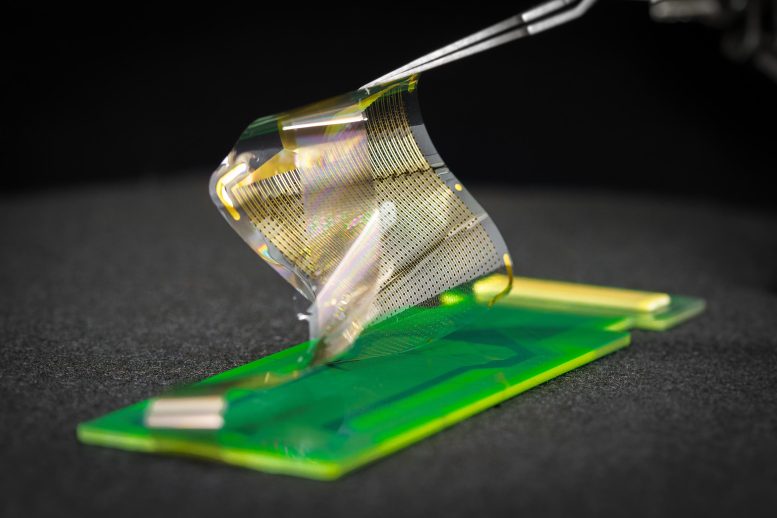
A new range of brain sensors can record electrical signals directly from the surface area of the human brain in record-breaking information. The new brain sensors feature thin, versatile, and largely packed grids of either 1,024 or 2,048 embedded electrocorticography (ECoG) sensors. Credit: David Baillot/ UC San Diego Jacobs School of Engineering
High-resolution recordings of electrical signals from the surface area of the brain could enhance surgeons ability to eliminate brain growths and deal with epilepsy, and could open brand-new possibilities for medium- and longer- term brain-computer interfaces.
A group of engineers, surgeons, and medical scientists has actually released information from both rats and people showing that a new array of brain sensors can record electrical signals straight from the surface area of the human brain in record-breaking detail. The brand-new brain sensors feature largely packed grids of either 1,024 or 2,048 ingrained electrocorticography (ECoG) sensors. The paper was published by the journal Science Translational Medicine on January 19, 2022.
These thin, pliable grids of ECoG sensors, if authorized for clinical use, would offer cosmetic surgeons brain-signal info straight from the surface of the brains cortex in 100 times greater resolution than what is readily available today. Access to this extremely in-depth perspective on which particular locations of the tissue at the brains surface area, or cortex, are active, and when, might provide better assistance for planning surgical treatments to remove brain growths and surgically treat drug-resistant epilepsy.
A brand-new range of brain sensors can tape electrical signals straight from the surface area of the human brain in record-breaking detail. A team of engineers, cosmetic surgeons, and medical researchers has published data from both rats and humans showing that a brand-new array of brain sensors can tape-record electrical signals directly from the surface of the human brain in record-breaking information. Recording brain activity from grids of sensing units put straight on the surface area of the brain– electrocorticography (ECoG)– is currently in common usage as a tool by surgeons carrying out treatments to remove brain tumors and deal with epilepsy in people who do not react to drugs or other treatments. Being able to tape-record brain signals at such high resolution might improve surgeons ability to get rid of as much of a brain growth as possible while reducing damage to healthy brain tissue. A new selection of brain sensors can tape electrical signals straight from the surface of the human brain in record-breaking information.
Pliable and thin grids of sensors in numerous sizes
The human brain is constantly moving. With each heartbeat, for example, the brain moves with the pulsating blood flowing through it. The platinum nano-rod based sensing unit grids are thinner and more versatile than todays medically approved ECoG grids. The thinness and flexibility permits the sensing unit grids to move with the brain, making it possible for a more detailed connection and better readings. In addition, the grids are produced with little, ring-shaped holes that enable cerebral spinal fluid to go through. In this method, these perfusion holes support a much better interface between the sensor grid and the brain surface area by allowing the sensor to easily and safely displace the fluid.
The brand-new platinum nano-rod brain sensing unit grids are ten micrometers thick, approximately one tenth the size of a human hair and 100 times thinner than the one millimeter thick and medically authorized ECoG grids. The nano-rods are embedded in a transparent, soft and versatile biocompatible material called parylene which is in direct contact with the surface area of the brain. The electrical signals travel from the brain, through the cerebrospinal fluid, and reach the exposed surface areas of the platinum nano-rods which are recessed within the parylene. This style yields a sensing unit grid that forms a close and steady connection with the surface area of the brain, enhancing signal quality.
The production process utilized also enables a wide range of shapes and sizes, which opens up new possibilities for greater and more customized cortical coverage. Gathering signals from larger areas of the brain at the very same time might unlock more of the brains secrets.
Customized sensor grids can be printed with specialized holes that allow surgeons to place probes at precisely the best area and apply electrical stimulation straight to brain tissue at specific locations. A new selection of brain sensors can record electrical signals straight from the surface of the human brain in record-breaking detail. The new brain sensing units feature thin, versatile, and densely packed grids of either 1,024 or 2,048 ingrained electrocorticography (ECoG) sensors.
More exact practical mapping
One of the difficulties of getting rid of brain tumors is that the presence of the growth triggers modifications in the brain, consisting of altering what areas of the brain are involved in what functions. These changes make it crucial for the surgical team to make a customized map of the patients brain– “practical maps”– in order to decide where to cut and where not to cut while removing as much of the tumor as possible.
The authors of the Science Translational Medicine paper demonstrated that these practical maps can be made exceptionally precise using their platinum nano-rod ECoG sensing units. In specific, the team developed functional maps in 4 different people of a boundary in the brain called the main sulcus. The main sulcus divides the brains somatomotor cortex from the somatosensory cortex. In these four individuals, the scientists positioned the platinum nano-rod grids on the surfaces of the topics brains and inquired to do a number of activities, including hand comprehending. With this information, the scientists rebuilded the actual place of this crucial landmark in the brain in addition to the neural associates in the brain that correspond to finger feeling and hand understanding. The results from the platinum nano-rod grids aligned with the arise from the lower-resolution ECoG grids already authorized for medical use, but with more precision about exactly where this important practical border is in between the somatomotor cortex and the somatosensory cortex. The freshly marked curvilinear practical limit distinct to each patients brain is superior to the often extrapolated and direct boundary that is determined from todays one-centimeter-spaced medical grids.
Neuroscience insights
In the past, the mapping of cortical columns has only been done by placement of a private needle at the surface of the brain and sequential electrical stimulation and movement of the needle throughout the brain surface area. More usually, the fact that the platinum nano-rod grids provide high resolution information in both time and area opens up lots of brand-new possibilities for discovering new understanding about how the brain works.
Another observation allowed by the brand-new grids is discovering the brief and regional in addition to long and broad variety brain waves associated with brain function all at the exact same time. This high- time-varying and spatial (vibrant) image of the brain activity was recorded in numerous additional films connected with the Science Translational Medicine paper and was used to translate hand movement in new methods using brain wave patterns.
Next steps
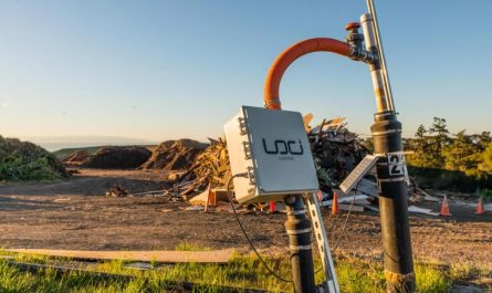
The group is dealing with a variety of initiatives in parallel to advance these grids so that they are eligible for evaluation for approval for brief-, medium- and longer-term usage. The team, led by UC San Diego electrical engineering professor Shadi Dayeh, was granted a $12.25 Million NIH grant focused on establishing the sensing system to the point that the next step will be a clinical trial for people with treatment-resistant epilepsy. This grant likewise funds efforts to make the system wireless, which would be essential for medium- and longer- term use.
Recommendation: “Human brain mapping with multithousand-channel PtNRGrids solves spatiotemporal dynamics” 19 January 2022, Science Translational Medicine.
Partnerships
The project is led by electrical engineering teacher Shadi Dayeh at the University of California San Diego Jacobs School of Engineering. The team of engineers, surgeons and medical researchers hails from UC San Diego; Massachusetts General Hospital; and Oregon Health & & Science University, where the bulk of the human surgeries were carried out by neurosurgery professor Ahmed Raslan. In addition to the Dayeh-Raslan cooperation, the development of the grids has covered several years of partnership between Dayeh and scientific partners spearheaded by Daniel Cleary of UC San Diego, and by Angelique Paulk and Jimmy Yang of Massachusetts General Hospital, and under the guidance of Sharona Ben-Haim and Eric Halgren of UC San Diego and Sydney Cash of Massachusetts General Hospital, and by Brittany Stedelin of Oregon Health & & Science University under Raslans supervision. Scaling of the grids and their interconnects were led by Dayehs postdoctoral fellow Youngbin Tchoe and college student Andrew Bourhis who is co-advised by Dayeh and Ian Galton of UC San Diego.
Innovation transfer
UC San Diego has actually submitted multiple patent applications on the manufacture of the multi-thousand channel PtNRGrids. Shadi Dayeh, Ahmed Raslan, and Youngbin Tchoe co-founded Precision Neurotech Inc. to support the commercialization of the PtNRGrids.
Funding
This work was supported by the National Institutes of Health Awards No. NBIB DP2-EB029757, NINDS R01NS123655-01, NINDS UG3NS123723-01 and NIDA R01-DA050159 and the National Science Foundation (NSF) Awards No. 1728497 and CAREER No. 1351980 to S.A.D., F32 postdoctoral fellowship to D.C. award # MH120886-01, and an NSF Graduate Research Fellowship Program No. DGE-1650112 to A.M.B.
Authors and Institutions
Co-First authors
Youngbin Tchoe, Andrew M. Bourhis, and Daniel R. Cleary from the Integrated Electronics and Biointerfaces Laboratory, Department of Electrical and Computer Engineering at the University of California San Diego Jacobs School of Engineering. Daniel Cleary is an MD/PhD whose primary appointment remains in the Department of Neurological Surgery at UC San Diego Health.
Corresponding author
Shadi A. Dayeh, who leads the Integrated Electronics and Biointerfaces Laboratory, Department of Electrical and Computer Engineering at the University of California San Diego Jacobs School of Engineering
Full Article Author List (by organization).
University of California San DiegoYoungbin Tchoe, Andrew M. Bourhis, Daniel R. Cleary, Jihwan Lee, Karen J. Tonsfeldt, Hongseok Oh, Yun Goo Ro, Keundong Lee, Samantha Russman, Mehran Ganji, Ian Galton, Sharona Ben-Haim, and Shadi A. Dayeh.
Oregon Health & & Science UniversityBrittany Stedelin, Erik C. Brown, Dominic Siler and Ahmed M. Raslan.
Massachusetts General HospitalAngelique C. Paulk, and Jimmy C. Yang.
Longer term, the group is working on wireless variations of these high resolution ECoG grids that might be utilized for up to 30 days of brain tracking for individuals with intractable epilepsy. The technology also holds possible for long-term implantation to improve the lifestyle of people who cope with paralysis or other neurodegenerative diseases that can be treated with electrical stimulation such as in Parkinsons illness, vital trembling, and the neurological motion condition called dystonia.
The task is led by electrical engineering professor Shadi Dayeh at the University of California San Diego Jacobs School of Engineering. The team of engineers, surgeons and medical scientists hails from UC San Diego; Massachusetts General Hospital; and Oregon Health & & Science University.
Animation illustrates use cases of the high-resolution brain sensing units described in the Science Translational Medicine paper entitled, “Human brain mapping with multithousand-channel PtNRGrids solves spatiotemporal dynamics” published on January 19, 2022. Video plays twice, as soon as with text labels and as soon as with just a clean animation. Credit: Angelique Paulk, Ahmed Raslan, Daniel Cleary, Youngbin Tchoe, and Shadi Dayeh
Next-generation electrocorticography
Recording brain activity from grids of sensing units put straight on the surface area of the brain– electrocorticography (ECoG)– is currently in typical usage as a tool by surgeons carrying out procedures to remove brain growths and deal with epilepsy in people who do not react to drugs or other treatments. The brand-new work in Science Translational Medicine supplies a large range of peer-reviewed data demonstrating that grids with 1,024 or 2,048 sensing units can be used to reliably tape and process electrical signals directly from the surface area of the brain in both rats and human beings. For comparison, the ECoG grids most commonly used in surgeries today usually have in between 16 and 64 sensors, although research grade grids with 256 sensors can be custom made.
Being able to tape brain signals at such high resolution might improve cosmetic surgeons capability to remove as much of a brain tumor as possible while minimizing damage to healthy brain tissue. In the case of epilepsy, higher resolution brain-signal recording capability might enhance a surgeons capability to exactly determine the regions of the brain where the epileptic seizures are stemming, so that these areas can be gotten rid of without touching close-by brain areas not associated with seizure initiation. In this way, these high resolution grids might enhance preservation of regular, working brain tissue.
Demonstrating that ECoG grids with sensing units in the thousands function well likewise opens new opportunities in neuroscience for revealing a deeper understanding of how the human brain functions. Fundamental science advances, in turn, could cause enhanced treatments grounded in boosted understanding of brain function.
A new selection of brain sensors can record electrical signals straight from the surface area of the human brain in record-breaking information. The brand-new brain sensing units include thin, flexible, and densely jam-packed grids of either 1,024 or 2,048 ingrained electrocorticography (ECoG) sensing units. Credit: David Baillot/ UC San Diego Jacobs School of Engineering
One millimeter vs one centimeter spacing
Recording brain signals at greater resolution is attributable to the groups ability to position private sensors substantially closer to each other without creating problematic disturbance in between nearby sensing units. The teams three-centimeter-by-three-centimeter grid with 1,024-sensors recorded signals directly from the brain tissue of 19 people who agreed to participate in this task throughout the “downtime” of their currently set up brain surgeries related to either cancer or epilepsy.
Sensing units made with platinum nano-rods
While using platinum-based sensors for recording electrical activity from neurons in the brain is not brand-new, the research study group is using platinum in an unique way: nanoscale platinum rods. The nano-rod shape offers more noticing surface location than flat platinum sensors, which assists to make the sensors more delicate. The sensing system is based upon the truth that the electron count in the platinum nano-rods changes in response to nerve cells shooting in the brain.
Charged ions move in and out of a neuron when it fires. This motion of charged ions causes modifications in the voltage capacity in the cerebrospinal fluid that the neurons are bathed in. These modifications in voltage capacity in the brain tissue and cerebrospinal fluid change the population count of electrons in the platinum nano-rods by charge screening procedures. In this method, the firing of nerve cells at or near the surface area of the cortex is tape-recorded by the platinum nanorods in near real time and with high precision.