The CXR extent of pneumonia was scored as 0 (no evidence of pneumonia). (B) Axial chest CT image at the lower lobe level (gotten on the same day) revealing negatively for pneumonia; CT level of pneumonia was scored as 0 (no proof of pneumonia). The CXR level of pneumonia was scored as 0 (no proof of pneumonia). CXR degree of pneumonia was scored as 0 (no evidence of pneumonia). Of patients undergoing CT, the proportion of CT scans without pneumonia was 22% (71/326) of unvaccinated clients, 30% (19/64) of partly vaccinated clients, and 59% (13/22) of totally immunized clients.
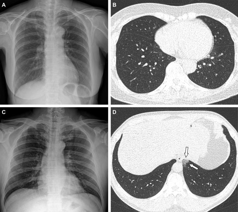
Representative cases showing pneumonia extents and patterns on chest X-ray (CXR) and CT images. (A and B) A 65-year-old female with development infection 2 months after a second dosage of the Pfizer vaccine (totally vaccinated). The client had a history of hypertension. (A) CXR acquired at admission showing no irregular opacification in both lung zones. The CXR level of pneumonia was scored as 0 (no evidence of pneumonia). (B) Axial chest CT image at the lower lobe level (acquired on the exact same day) showing negatively for pneumonia; CT degree of pneumonia was scored as 0 (no evidence of pneumonia). (C and D) A 48-year-old male with 1 month after a very first dose of the AstraZeneca vaccine (partly immunized). The patient had no history of comorbidity. (C) CXR gotten at admission showing no abnormal opacification in both lung zones. The CXR degree of pneumonia was scored as 0 (no evidence of pneumonia). (D) Axial chest CT image acquired on the same day revealing unilateral ground-glass opacity with a non-rounded morphology in the left lower lobe (arrows). CT level of pneumonia was scored as 1 (1-25% involvement) and this case was classified as indeterminate look of COVID-19 according to the RSNA chest CT category system. Credit: Radiological Society of North America
The clinical and imaging qualities of COVID-19 advancement infections in fully immunized clients tend to be milder than those of partially vaccinated or unvaccinated clients, according to a new multicenter research study published in the journal Radiology.
The variety of verified COVID-19 cases worldwide now goes beyond 270 million with a total mortality rate of approximately 2%.
COVID-19 vaccines are important and efficient tools for bringing the pandemic under control. Vaccines are not 100% efficient at preventing illness. Development infections are specified as the detection of serious intense respiratory syndrome coronavirus 2 (SARS-CoV-2) ribonucleic acid (RNA) or antigen in a breathing specimen gathered from a person 14 days or more after getting all recommended doses of COVID-19 vaccines.
Advancement cases are on the increase with the extremely transmissible Omicron variation. For that reason, it is very important to understand how vaccination impacts not just COVID-19 disease seriousness but likewise medical information and medical imaging results.
” Although the risk of infection is much lower amongst immunized people, and vaccination minimizes the seriousness of disease, medical and imaging data of COVID-19 breakthrough infections have actually not been reported in information,” stated the research studys senior author, Yeon Joo Jeong, M.D., Ph.D., from the Department of Radiology and Biomedical Research Institute at Pusan National University Hospital in Busan, South Korea. “The purpose of this study was to record the clinical and imaging functions of COVID-19 breakthrough infections and compare them with those of infections in unvaccinated clients.”
Agent cases showing pneumonia extents and patterns on chest X-ray (CXRs) and CT images. (E and F) A 36-year-old male without any history of vaccination for COVID- 19. The patient had no history of comorbidity. (E) CXR acquired at admission showing no irregular opacification in either lung zone. CXR degree of pneumonia was scored as 0 (no evidence of pneumonia). (F) Axial chest CT image gotten on the very same day revealing unilateral ground-glass opacity with a nonrounded morphology and non-peripheral distribution in the left upper lobe (arrows). CT level of pneumonia was scored as 1 (1-25% participation) and this case was categorized as indeterminate look of COVID-19 according to the RSNA chest CT category system. (G and H) A 58-year-old male with no history of COVID-19 vaccination. The patient had a history of hypertension and diabetes. He required supplemental oxygen on admission and was confessed to intensive care unit one day later. (G) CXR at admission revealing patchy ground-glass opacities in both middle- to lower-lung zones. CXR level of pneumonia was scored as 2 (>> 25% participation). (H) Axial chest CT image gotten on the same day revealing multifocal ground-glass opacities with a crazy-paving look in bilateral lungs. CT degree of pneumonia was scored as 2 (>> 25% involvement) and was classified as normal look of COVID-19 according to the RSNA chest CT category system. Credit: Radiological Society of North America
In this retrospective multicenter friend study, Dr. Jeong and colleagues evaluated data from adult clients signed up in an open data repository for COVID-19– Korean Imaging Cohort for COVID-19 (KICC-19)– in between June and August 2021. Hospitalized clients with standard chest X-rays were divided into three groups, according to their vaccination status. The researchers examined distinctions in between clinical and imaging functions and evaluated associations between scientific factors– including vaccination status– and scientific outcomes.
Chest CT scans were carried out on 412 (54%) of the clients throughout hospitalization. Of patients going through CT, the proportion of CT scans without pneumonia was 22% (71/326) of unvaccinated patients, 30% (19/64) of partly immunized clients, and 59% (13/22) of fully vaccinated clients.
The results also revealed associations between the danger of severe disease and clinical attributes such as greater age, history of diabetes, lymphocytopenia, thrombocytopenia, elevated LDH (lactate dehydrogenase), and raised CRP (C-reactive protein). Notably, age was also found to be an important predictor of more extreme disease in COVID-19 patients, even in those with an advancement infection.
The researchers noted that observed differences in clinical attributes may reflect distinctions in vaccination concerns based on underlying comorbidities. Throughout the study period, high-risk groups, such as people over 65 years of ages, healthcare employees and individuals with impairments were concern targets for COVID-19 vaccination. Elderly clients and clients with at least one comorbidity were more common in the immunized group than in unvaccinated group in the study.
” Despite these differences, mechanical ventilation and in-hospital death took place only in the unvaccinated group,” Dr. Jeong said. “Furthermore, after changing for baseline medical qualities, analysis showed that totally immunized clients were at significantly lower risk of needing supplemental oxygen and of ICU admission than unvaccinated clients.”
Additional research will be required as different versions emerge, this study sheds light on the clinical efficiency of COVID-19 vaccination in the context of advancement infections.
Recommendation: “Imaging and Clinical Features of COVID-19 Breakthrough Infections: A Multicenter Study” 1 February 2022, Radiology.
Collaborating with Dr. Jeong were Jong Eun Lee, M.D., Minhee Hwang, M.D., Yun-Hyeon Kim, M.D., Ph.D., Myung Jin Chung, M.D., Ph.D., Byeong Hak Sim, M.D., Kum Ju Chae, M.D., and Jin Young Yoo, M.D.


