In Bultes experience, he states, identifying healing cells that are prepared for human clinical trials in any organ is a tough and expensive process, needing comprehensive security studies and keeping teams of scientists focused on finding much better ways to track restorative cells damaged by several sclerosis and other neurodegenerative diseases.
For such research, researchers have actually long been utilizing so-called mesenchymal stromal cells, a kind of stem cell discovered in the bone marrow that can become many types of cells, and also reduce swelling.
In the new proof-of principle research study explained today (February 7, 2022) in Nature Biomedical Engineering, Bulte and his research study team discovered that these mesenchymal stromal cells consist of high levels of a sugar called mannose, which is similar to glucose and can be spotted quickly and effectively with a basic imaging technique based upon magnetic resonance imaging (MRI).
Bultes group understood to utilize sugars as tracers after they published another study in Nature Communications revealing that specific tumor cells lose big amounts of sugar particles easily discovered by MRI devices. Yue Yuan, Ph.D., a researcher in Bultes lab, discovered that stem cells have an abundance of mannose– about 2 to 3 times the amount discovered in common cells.
Due to the fact that the cells of mammals typically lack a high sugar material, Bulte and his group reasoned, injections of stem cells that naturally include lots of sugar would potentially be easy to spot against the background of brain tissue.
For their study, Bultes team injected four kinds of human cells into the brains of live mice, consisting of mesenchymal stem cells. For each cell type, the scientists injected 300,000 cells.
The researchers utilized MRI to track where they discovered clusters of the injected cells over a two-week duration.
They discovered that the MRI signal of mesenchymal stem cells was about 60% greater than the other 3 injected cell types, and was easily seen on MR images up to two weeks after injection. Surprisingly, they state, just live cells produced an MRI signal, producing a chance to utilize the method for identifying transplanted cell survival, in addition to tracking.
If the cellular sugar molecules can be utilized to identify distinction of stem cells into other cell types, Bultes team is planning additional studies to identify.
” Its extraordinary to find, 30 years after beginning my research study in the field of labeling cells, that these mesenchymal stem cells in the brain do not require to be chemically identified for tracking purposes after all, and there might be better, easier ways to track these cells in the brain,” states Bulte.
Reference: 7 February 2022, Nature Biomedical Engineering.DOI: 10.1038/ s41551-021-00822-w.
This research study was supported by the Pearl and Yueh-Heng Yang Foundation and the National Institutes of Health (R56 NS098520 and P41 EB024495).
In addition to Bulte and Yuan, scientists who added to the research while at Johns Hopkins include Congxiao Wang, Shreyas Kuddannaya, Jia Zhang, Dian R. Arifin, Zheng Han, Piotr Walczak and Guanshu Liu.
Bulte is a paid specialist to NovaDip Biosciences SA, NanomediGene LLC, and SuperBranche. These plans have been evaluated and approved by The Johns Hopkins University in accordance with its conflict-of-interest policies.
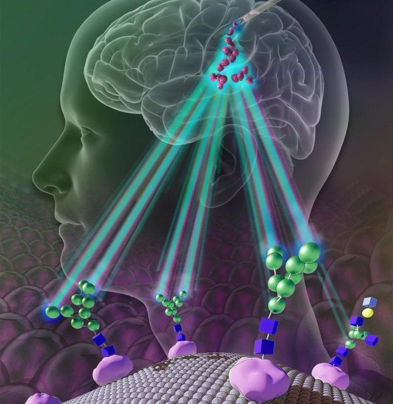
Unlike most other cells, mannose residues (green) are abundant on the MSC cell membrane. Using an MRI method that is sensitive to the existence of mannose, tracking of transplanted MSCs is now possible without the requirement of labeling them. Credit: Image courtesy of Shreyas Kuddannaya
A Johns Hopkins Medicine scientist who spent 30 years determining how to put chemical labels into cells to track their movement in living tissues has found that certain self-renewing stem cells have integrated tracers– constructed of sugars– that can do the task without added chemical “labels” when injected into mouse brains. The finding, made with stem cells extensively engineered into speculative treatments for multiple sclerosis and other neurodegenerative illness, was a welcome surprise, the investigators state.
” There is a whole scientific field devoted to chemical and genetic cell labeling, due to the fact that otherwise, we cant see where specially and expensively crafted healing cells travel and whether they get to the designated area in a body to repair or change infected tissue,” states Jeff Bulte, Ph.D., teacher of radiology and radiological science at the Johns Hopkins University School of Medicine and director of cell imaging for the Johns Hopkins Institute for Cell Engineering.
If confirmed with subsequent experiments, the new study should, Bulte states, simplify and advance corrective research for diseases of the brain, an organ considered the most difficult in which to track therapies due to the fact that of the delicate nature of the brain and its blood-brain barrier.