But amyloid deposits might likewise happen in the retina of the eye, typically in patients clinically diagnosed with AD, suggesting similar pathologies in both organs. In a small, cross-sectional research study, a team of scientists, led by researchers at University of California San Diego School of Medicine, compared tests of retinal and brain amyloids in clients from the A4 study and another study (Longitudinal Evaluation of Amyloid Risk and Neurodegeneration) assessing neurodegeneration danger personallies with low levels of amyloid.
Like the proverbial “windows to the soul,” the researchers observed that the presence of retinal areas in the eyes correlated with brain scans showing higher levels of cerebral amyloid. The finding recommends that non-invasive retinal imaging might work as a biomarker for finding early-stage advertisement threat.
The findings were published in the August 17, 2021 problem of Alzheimers & & Dementia
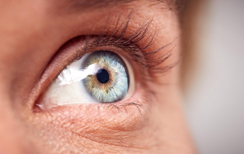
. Amyloid deposits tagged by curcumin fluoresce in a retinal scan. Credit: NeuroVision
” This was a little preliminary dataset from the screening check out. It included eight patients,” stated senior author Robert Rissman, PhD, teacher of neurosciences at UC San Diego School of Medicine and director of the Biomarker Core for the Alzheimers Disease Cooperative Study and the Shiley-Marcos Alzheimers Disease Research Center at UC San Diego. “But these findings are motivating since they recommend it may be possible to figure out the beginning, spread and morphology of advertisement– a preclinical diagnosis– using retinal imaging, instead of more difficult and pricey brain scans. We anticipate seeing the outcomes of additional timepoint retinal scans and the impact of solanezumab (a monoclonal antibody) on retinal imaging. Unfortunately, we will need to wait to see and evaluate these information when the A4 trial is completed.”
The next step, stated Rissman, will be to perform a bigger study to more completely document and determine the relationship between retinal amyloid and cerebral amyloid, both cross-sectionally and with time.
Reference: “Feasibility study for detection of retinal amyloid in medical trials: The Anti-Amyloid Treatment in Asymptomatic Alzheimers Disease (A4) trial” by Jennifer Ngolab, Michael Donohue, Alison Belsha, Jennifer Salazar, Paula Cohen, Sandhya Jaiswal, Veasna Tan, Devon Gessert, Shaina Korouri, Neelum T. Aggarwal, Jessica Alber, Ken Johnson, Gregory Jicha, Christopher van Dyck, James Lah, Stephen Salloway, Reisa A. Sperling, Paul S. Aisen, Michael S. Rafii and Robert A. Rissman, 17 August 2021, Alzheimers & & Dementia.DOI: 10.1002/ dad2.12199.
Co-authors include: Jennifer Ngolab and Shaina Korouri, UC San Diego; Michael Donohue, Alison Belsha, Jennifer Salazar, Paula Cohen, Sandhya Jaiswal, Veasna Tan, Devon Gessert, Paul S. Aisen and Michael S. Rafii, all at University of Southern California; Neelum T. Aggarwal, Rush University Medical Center; Jessica Alber, University of Rhode Island; Ken Johnson, NeuroVision Imaging Inc; Gregory Jicha, University of Kentucky; Christopher van Dyck, Yale University; James Lah, Emory University; Stephen Salloway, Butler Hospital, R.I.; Reisa A. Sperling, Brigham and Womens Hospital/Massachusetts General Hospital, Boston.
Financing for this research came, in part, from the National Institutes of Health and National Institute on Aging (grants AG062429, AG018440, AG010483, AG063689), Alzheimers Association, Eli Lilly and Company, NeuroVision Inc., Accelerating Medicines Partnership, GHR Foundation, Morton Plant Mease Foundation, plus a confidential structure and additional personal donors.
Amyloid deposits tagged by curcumin fluoresce in a retinal scan. “But these findings are encouraging because they suggest it may be possible to identify the beginning, spread and morphology of AD– a preclinical diagnosis– using retinal imaging, rather than more difficult and costly brain scans. We look forward to seeing the outcomes of additional timepoint retinal scans and the effect of solanezumab (a monoclonal antibody) on retinal imaging.
Protein deposits in retina and brain appear to parallel possible neurodegeneration, an insight that might result in simpler, quicker detection.
Amyloid plaques are protein deposits that gather in between brain cells, preventing function and eventually leading to neuronal death. They are considered a trademark of Alzheimers disease (ADVERTISEMENT), and the focus of numerous investigations developed to lower or prevent their development, consisting of the nationwide A4 study.