The human body consists of around 37 trillion cells; the fruit fly sweeping around the overripe bananas on your counter may have 50,000 cells. These genes, when placed into a cells genome, trigger it to produce microscopic hollow protein structures understood as gas blisters. Shapiro says recent work on engineered stress of injectable germs that attack cancer cells, or “tumor-homing” germs, produces a need for better methods to track these cells to see where in the body they land. “There are some cases where single bacterial cells that are extremely little and have an extremely large amount of these gas blisters are damaged, however it doesnt make much of a distinction to the bacterial population if a few of them end up being less feasible. That will involve engineering and screening brand-new kinds of blisters that have different residential or commercial properties, such as blisters that pop more quickly, or vesicles that are more robust, or smaller sized vesicles that can fit into locations that bigger blisters can not.
These fluorescent reporter genes have a huge disadvantage though: light does not penetrate really far through living tissues.
So, Shapiro has actually developed reporter genes that use sound rather of light. These genes, when inserted into a cells genome, cause it to produce tiny hollow protein structures referred to as gas blisters. These blisters are usually found in specific types of germs that utilize them to survive in water, but they likewise have the beneficial residential or commercial property of “sounding” when struck by ultrasound waves.
The idea is that when a cell producing these blisters is imaged with ultrasound, it will send an acoustic signal announcing its presence, enabling researchers to see where it is and what it is doing. This technique has been utilized to reveal the activity of enzymes in cells in previous work by Shapiros laboratory.
In their most current paper, the research group explains how it has increased the sensitivity of that method so much that it can now image a single cell, located within body tissue, that is bring an acoustic press reporter gene.
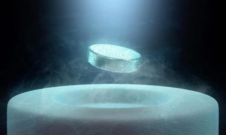
Single cells taking a trip through the liver of a mouse are highlighted by a brand-new imaging method established in Mikhail Shapiros laboratory. Credit: Caltech/Daniel Sawyer, Shapiro Lab
” In comparison to previous work on gas blisters, this paper enables us to see much smaller amounts of these gas vesicles,” says Daniel Sawyer (PhD 21), lead author and previous bioengineering PhD student in Shapiros lab. “This is like going from a satellite that can see the lights of a village to one that can see the light from a single lamppost.”
Their improvements represent an increase of more than 1000-fold in sensitivity over the previous method they had actually been using for imaging cells bring the acoustic press reporter genes. The difference depends on the ultrasound they utilize and how the gas vesicles react to it.
Whereas the previous imaging strategy depended on the vesicles calling like a bell that has actually been struck, the new strategy uses stronger ultrasound that “pops” the vesicles like a balloon.
” The blisters produce a very strong signal because minute,” Shapiro states. “Then the vesicles break and stop making a signal. Were looking for the little blip.”
That blip is so clear that it can easily be detected by the researchers, even amidst all the background sound produced by ultrasound permeating through tissue. Shapiro says recent deal with engineered strains of injectable bacteria that attack cancer cells, or “tumor-homing” germs, creates a requirement for much better ways to track these cells to see where in the body they land. The researchers revealed that when the germs were likewise engineered to carry the gas-vesicle gene, it was possible to track private bacterial cells as they went into and took a trip through the liver after being injected into the blood stream.
Sawyer says this level of sensitivity is required if researchers wish to use ultrasound for studying the structure of the gut microbiome, which, when interfered with, can affect conditions like Alzheimers illness and autism.
” There are numerous types of germs in your gut, and some are so rare that you need something delicate enough to see simply the few of them deep inside the body,” he says.
Does popping the blisters inside cells damage the cells? No, not really.
” The brief answer is no, and the long response is no in a lot of practical cases,” Sawyer states. “There are some cases where single bacterial cells that are really small and have a large amount of these gas vesicles are damaged, however it doesnt make much of a distinction to the bacterial population if a few of them become less feasible. And in mammalian cells, we saw no unfavorable result.”
Shapiro and Sawyer are pursuing 2 paths for their research going forward. That will involve engineering and screening brand-new kinds of vesicles that have different residential or commercial properties, such as blisters that pop more quickly, or vesicles that are more robust, or smaller sized vesicles that can fit into locations that larger vesicles can not.
,” Shapiro says. “Dannys paper is part of the advancement of the ultrasound analogue of those imaging methods.”
The paper describing their research study, entitled, “Ultrasensitive ultrasound imaging of gene expression with signal unmixing,” appears in the August 6 concern of the journal Nature Methods. Co-authors include Avinoam Bar Zion, visitor in chemical engineering; Arash Farhadi (PhD 20); bioengineering college student Shirin Shivaei; chemical engineering graduate trainee Bill Ling; and Audrey Lee-Gosselin, previously of Caltech.
Reference: “Ultrasensitive ultrasound imaging of gene expression with signal unmixing” by Daniel P. Sawyer, Avinoam Bar-Zion, Arash Farhadi, Shirin Shivaei, Bill Ling, Audrey Lee-Gosselin and Mikhail G. Shapiro, 5 August 2021, Nature Methods.DOI: 10.1038/ s41592-021-01229-w.
Financing for the research was offered by the National Institutes of Health.
Mikhail Shapiro is an affiliated professor of the Tianqiao and Chrissy Chen Institute for Neuroscience.
Credit: Barth van Rossum for Caltech
If you are a researcher who desires to see how just a couple of cells in an organism are acting, it is no basic job. The human body contains around 37 trillion cells; the fruit fly sweeping around the overripe bananas on your counter may have 50,000 cells.
Researchers working in the Caltech laboratory of Mikhail G. Shapiro, professor of chemical engineering and Heritage Medical Research Institute Investigator, have found a method.
The brand-new strategy utilizes so-called acoustic reporter genes, of which Shapiro has actually been a pioneering designer. To understand acoustic press reporter genes, first understand that press reporter genes are a specialized bit of DNA that scientists can place into an organisms genome to help them comprehend what it is doing. Historically, press reporter genes have actually encoded fluorescent proteins. If a researcher inserts one of these reporter genes next to a gene they desire to study– say, the gene that is accountable for the development of neurons– the activation of those nerve cell genes will also produce fluorescent protein particles. When the right sort of light is shined upon those cells, they will illuminate, type of like how a highlighter can mark a particular passage in a book.