
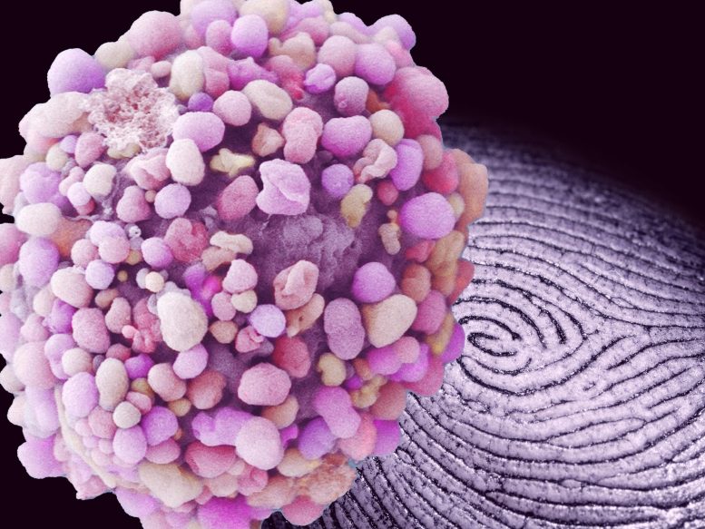
MIT researchers have established a method to decode images of cells to determine whether a tissue is more like a strong, liquid, or even a gas. These visual “fingerprints” might help to rapidly identify and track different cancers. Credit: breast cancer cell by Anne Weston, Francis Crick Institute, modified by MIT News
The technique might be a path to quicker, less intrusive cancer diagnoses.
In the preliminary phases, an embryo takes on a practically fluid-like state that permits its cells to broaden and divide. As it matures, its tissues and organs company up into their last form.
Now, scientists at MIT have actually discovered that the method in which a tissues cells are set up can serve as a fingerprint for the tissues “stage.” They have developed a method to decode pictures of cells in a tissue to quickly determine whether that tissue is more like a solid, liquid, or perhaps a gas. Their findings were just recently reported in the Proceedings of the National Academy of Sciences.
MIT scientists have established a way to decipher images of cells to determine whether a tissue is more like a strong, liquid, or even a gas. Credit: breast cancer cell by Anne Weston, Francis Crick Institute, edited by MIT News
In the preliminary stages, an embryo takes on a nearly fluid-like state that enables its cells to divide and broaden. They have actually established an approach to decipher images of cells in a tissue to quickly identify whether that tissue is more like a solid, liquid, or even a gas. The scientists then went on to use their technique in systems with cells rather than molecules.
The team hopes that their approach, which theyve dubbed “configurational fingerprinting,” can help researchers track physical changes in an embryo as it develops. More right away, they are using their technique to study and ultimately diagnose a specific type of tissue: tumors.
In cancer, there has been proof to recommend that, like an embryo, a tumors physical state may suggest its stage of development. Growths that are more strong may be relatively stable, whereas more fluid-like growths might be more susceptible to alter and metastasize.
The MIT researchers are examining images of growths, both grown in the laboratory and biopsied from clients, to determine cellular fingerprints that show whether a tumor is more like a strong, liquid, or gas. They visualize that medical professionals can one day match a picture of a tumors cells with a cellular fingerprint to rapidly identify a growths stage, and eventually a cancers development.
” Our technique would permit a really simple medical diagnosis of the states of cancer, just by examining the positions of cells in a biopsy,” says Ming Guo, associate professor of mechanical engineering at MIT. “We hope that, by simply looking at where the cells are, doctors can directly tell if a tumor is really solid, indicating it cant metastasize yet, or if its more fluid-like, and a client is in risk.”
Guos co-authors are Haiqian Yang, Yulong Han, Wenhui Tang, and Rohan Abeyaratne of MIT, Adrian Pegoraro of the University of Ottawa, and Dapeng Bi of Northeastern University.
Triangular order
In a best solid, the products private constituents are configured as an orderly lattice, such as the atoms in a cube of crystal. If you were to cut a slice of the crystal and lay it on a table, you would see that the atoms are organized such that you could connect them in a pattern of duplicating triangles. In a perfect strong, as the spacing between atoms would be precisely the very same, the triangles that connect them would generally be equilateral fit.
Guo took this construct as a template for a perfectly strong structure, with the concept that it might act as a reference for comparing the cell setups of actual, less-than-perfectly-solid tissues and tumors.
” Real tissues are never completely purchased,” Guo says. “They are mostly disordered. But still, there are subtle distinctions in just how much they are disordered.”
Following this idea, the group started with images of different types of tissues and utilized software to map triangular connections between a tissues cells. In contrast to the equilateral triangles in a best solid, the maps produced triangles of numerous shapes and sizes, showing cells with a variety of spatial order (and condition).
For each triangle in an image, they determined 2 key criteria: volumetric order, or the space within a triangle; and shear order, or how far a triangles shape is from equilateral. The very first criterion indicates a materials density variation, while the second shows how vulnerable the material is to warping. These two parameters, they discovered, sufficed to identify whether a tissue was more like a solid, liquid, or gas.
” Were directly computing the precise value of both parameters, compared to those of a perfect strong, and utilizing those precise worths as our finger prints,” Guo describes.
Vapor tendrils
For each concentration, they mapped the molecules into triangles, then measured each triangles 2 criteria. From these measurements, they characterized the stage of the molecules and were able to reproduce the transitions between gas, liquid, and strong, that was expected.
” People know what to expect in this really basic system, and this is what we see exactly,” Guo says. “This showed the ability of our approach.”
The scientists then went on to use their approach in systems with cells rather than particles. For instance, they looked at videos, taken by other scientists, of a growing fruitfly wing. Applying their technique, they might identify areas in the establishing wing that changed from strong to a more fluid state.
” As a fluid, this may aid with growth,” Guo states. “How precisely that happens is still under investigation.”
He and his group likewise grew small tumors from cells of human breast tissue and watched as the tumors grew appendage-like tendrils– indications of early transition. When they mapped the setup of cells in the growths, they discovered that the noninvasive tumors resembled something between a strong and a liquid, and the invasive tumors were more gas-like, while the tendrils showed a lot more disordered state..
” Invasive growths were more like vapor, and they wish to spread out and go all over,” Guo states. “Liquids can hardly be compressed. However gases are compressible– they can swell and diminish quickly, and thats what we see here.”.
The team is working with samples of human cancer biopsies, which they are imaging and examining to develop their cellular fingerprints. Ultimately, Guo envisions that mapping a tissues phases can be a fast and less invasive method to detect numerous kinds of cancer.
” Doctors normally need to take biopsies, then stain for various markers depending upon the cancer type, to detect,” Guo says. “Perhaps one day we can use optical tools to look inside the body, without touching the client, to see the position of cells, and straight inform what stage of cancer a patient is in”.
Referral: “Configurational finger prints of multicellular living systems” by Haiqian Yang, Adrian F. Pegoraro, Yulong Han, Wenhui Tang, Rohan Abeyaratne, Dapeng Bi, and Ming Guo, 25 October 2021, Proceedings of the National Academy of Sciences.DOI: 10.1073/ pnas.2109168118.
This research study was supported in part by the National Institutes of Health, MathWorks, and the Jeptha H. and Emily V. Wade Award at MIT.