
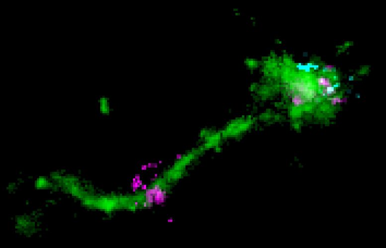
An LNP is located on a long endosomal tubule (green), together with a perpendicular disperse mRNA signal (cyan), and likely representing an instance of mRNA (purple) escape. Credit: Marino Zerial/ MPI-CBG
Scientists have actually discovered where and how mRNA gets here in a cell to modify or deliver hereditary information, a crucial process for the development of unique treatments.
DNA (deoxyribonucleic acid) contains the hereditary details needed for the development and maintenance of life. This information is communicated by messenger ribonucleic acid (mRNA) to make proteins. mRNA-based therapies have the prospective to resolve unmet needs for a wide array of illness, consisting of cancer and cardiovascular illness. mRNA can be delivered to cells to activate the production, degradation, or modification of a target protein, something impossible with other approaches.
A crucial challenge with this technique is having the ability to deliver the mRNA inside the cell so that it can be translated to make a protein. mRNA can be packed into lipid nanoparticles (LNPs)– little bubbles of fat– that secure the mRNA and shuttle it into cells. However, this process is not simple, since the mRNA needs to pass the membrane prior to it can reach its website of action in the cell interior, the cytoplasm.
A crucial difficulty with this technique is being able to deliver the mRNA inside the cell so that it can be translated to make a protein. Researchers in the team of MPI-CBG director Marino Zerial are experts in visualizing the cellular entry routes of molecules in the cell, such as mRNA with high-resolution microscopes. They teamed up with researchers from AstraZeneca who supplied the researchers with lipid nanoparticle prototypes that they had actually established for therapeutic techniques to follow the mRNA inside the cell. At this point, the mRNA is inside the cells however surrounded by two barriers, the fatty bubble and the endosome wall or more correctly, membrane.” With single particle microscopy methods,” discusses Prasath Paramasivam, the very first author of the research study, “we could picture for the first time the mRNA in the LNP inside the endosomes of cells.
Scientists in the group of MPI-CBG director Marino Zerial are professionals in picturing the cellular entry paths of particles in the cell, such as mRNA with high-resolution microscopic lens. They teamed up with scientists from AstraZeneca who supplied the scientists with lipid nanoparticle prototypes that they had actually developed for restorative approaches to follow the mRNA inside the cell. The study is released in the Journal of Cell Biology.
” To be provided, the mRNA should make a long journey. Confined in the fatty LNP bubble, it requires to get into the cell initially,” describes Marino Zerial. “The LNPs come to the cell surface where they bind to receptors. They are then taken up into specialized membrane-enclosed compartments called endosomes. At this point, the mRNA is inside the cells however surrounded by two barriers, the fatty bubble and the endosome wall or more correctly, membrane. The difficulty for the mRNA is to leave both barriers to reach the cytoplasm where it works as a template to make proteins. We understand that only a tiny portion of RNA molecules are able to leave into the cytoplasm.”
Internalized cargo molecules, like the LNPs, are first transported to “early” endosomes. These are logistic centers that distribute freight particles to numerous destinations in the cell. They either recycle particles to the cell surface or degrade them in late endosomes and lysosomes. Far, individuals thought that the mRNA gets away from late endosomes exploiting their extremely acidic material.
” With single molecule microscopy methods,” discusses Prasath Paramasivam, the first author of the research study, “we could envision for the first time the mRNA in the LNP inside the endosomes of cells. We likewise caught the actual escape of the mRNA, which took place in the tubules of the recycling endosomes, which are only mildly acidic. Our outcomes imply that sending out the LNP-mRNA to late endosomes is detrimental for shipment and only increases cell toxicity,” states Zerial. These findings assist in comprehending the mechanism of mRNA escape from endosomes in more information.
Marino Zerial summarizes: “The LNP shipment system for mRNA demands high doses due to the low endosomal escape efficiency. Understanding where the mRNA goes and how it can escape the endosomes allows us to develop better cars for more effective delivery, at lower dosage. We can improve the mRNA shipment system so it can be used for healing applications, for example, cancer treatment.”
Referral: “Endosomal escape of delivered mRNA from endosomal recycling tubules imagined at the nanoscale” by Prasath Paramasivam, Christian Franke, Martin Stöter, Andreas Höijer, Stefano Bartesaghi, Alan Sabirsh, Lennart Lindfors, Marianna Yanez Arteta, Anders Dahlén, Annette Bak, Shalini Andersson, Yannis Kalaidzidis, Marc Bickle and Marino Zerial, 9 December 2021, Journal of Cell Biology.DOI: 10.1083/ jcb.202110137.