Healthy heart tissue (left) and in severe Covid-19 (right). While the capillary in the healthy case show an undamaged structure (bottom right), one can see cavities or pouches and tiny tubular structures forming in the fine blood vessels (blood vessels) in the Covid-19 tissue sample, which suggest particular morphological modifications. Credit: M Reichardt, T Salditt
In contrast with a healthy heart, X-ray imaging of tissues impacted by severe disease, revealed a network full of divides, branches, and loops which had actually been chaotically remodeled by the formation and splitting of new vessels. These modifications are the very first direct visual proof of among the primary motorists of lung damage in Covid-19: a special kind of “intussusceptive angiogenes” (indicating brand-new vessel development) in the tissue.
“To speed up image processing, we therefore also automatically broke the tissue architecture down into its local balanced features and then compared them,” discusses Marius Reichardt, at the University of Göttingen and first author of the paper. “The criteria gotten from this then revealed an entirely various quality compared to healthy tissue, or even to illness such as serious influenza or typical myocarditis,” explain the leaders of the study, Professor Tim Salditt from the University of Göttingen and Professor Danny Jonigk from the MHH.
There is a really special function of this study: in contrast to the vascular architecture, the required data quality might be achieved utilizing a little X-ray source in the laboratory of the University of Göttingen. In concept, this implies it might also be performed in any center to support pathologists with routine diagnostics. In the future, the researchers wish to more broaden the method of transforming the particular tissue patterns into abstract mathematical values in order to establish automated tools for diagnostics, once again by more developing laboratory X-ray imaging and validating it with data from synchrotron radiation. The collaboration with DESY will be further broadened in the coming years.
Referral: “3D virtual histopathology of heart tissue from Covid-19 patients based on phase-contrast X-ray tomography” by Marius Reichardt, Patrick Moller Jensen, Vedrana Andersen Dahl, Anders Bjorholm Dahl, Maximilian Ackermann, Harshit Shah, Florian Länger, Christopher Werlein, Mark P Kuehnel, Danny Jonigk and Tim Salditt, 21 December 2021, eLife.DOI: 10.7554/ eLife.71359.
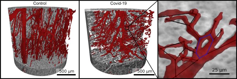
Vascular network (red) in healthy heart tissue (left) and in severe Covid-19 (right). An interdisciplinary research team from the University of Göttingen and Hannover Medical School (MHH) has actually found substantial modifications in the heart muscle tissue of individuals who passed away from Covid-19. They observed clear changes at the level of the capillaries (the tiny blood vessels) in the heart muscle tissue when they analyzed the results there of the severe form of Covid-19 illness.
Healthy heart tissue (left) and in severe Covid-19 (right).
Vascular network (red) in healthy heart tissue (left) and in extreme Covid-19 (right). Due to malfunctioning reforming of the network as a result of Covid-19, numerous branches, splits and even loops establish in the blood vessels, which can be examined mathematically. Credit: M. Reichardt, P. Møller Jensen, T. Salditt
Interdisciplinary research team from Göttingen University and Hannover Medical School are very first to show this straight.
An interdisciplinary research study group from the University of Göttingen and Hannover Medical School (MHH) has actually found considerable modifications in the heart muscle tissue of people who died from Covid-19. The existing study underpins the involvement of the heart in Covid-19 at the microscopic level for the first time by imaging and analyzing the affected tissue in the 3 measurements.
The scientists imaged the tissue architecture to a high resolution using synchrotron radiation– an especially intense X-ray radiation– and displayed it three-dimensionally. To do this, they used a special X-ray microscopic lense that the University of Göttingen set up and runs at the German Electron Synchrotron DESY in Hamburg. They observed clear modifications at the level of the capillaries (the small capillary) in the heart muscle tissue when they took a look at the results there of the severe type of Covid-19 disease.