
The results, reported by scientists at the University of California, Berkeley, and UC San Francisco (UCSF), offer a possible description for why some individuals are resistant and others susceptible to traumatic stress, and for the different symptoms– avoidance habits, stress and anxiety and worry, for instance– triggered by the memory of such tension.
If, as the scientists suspect, extreme injury causes the increased myelination, the findings could cause treatments– drugs or behavioral interventions– that avoid or reverse the myelin production and decrease the effects of severe injury.
Myelin is a layer of fatty compounds and proteins that covers around the axons of neurons– basically, the insulation around the brains circuitry– to assist in long-distance transmission of signals and, thus, communication between remote areas of the brain. The inner regions of the brain look white– in reality, they are referred to as “white matter”– due to the fact that of the myelin encasing the many big bundles of axons there.
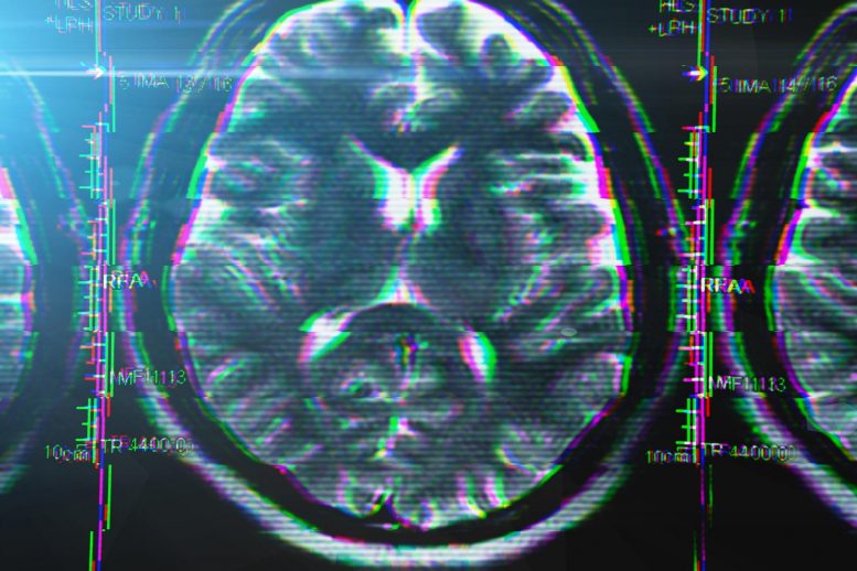
An fMRI scan of the brain of a military veteran with PTSD, revealing gray matter areas with increased myelin. Credit: UCSF image by Linda Chao
But the new study finds increased myelination of axons in so-called “noodle,” where most of the cell bodies of neurons live and most of the wiring is less insulated with myelin. The extra myelination was discovered mostly in areas connected with memory.
Scientists at the San Francisco Veterans Affairs Medical Center conducted brain MRI scans of 38 veterans– half with PTSD, half without– and discovered a boost in myelination in the gray matter of those with PTSD compared to that seen in the brains of those not struggling with PTSD.
Associates at UC Berkeley, meanwhile, discovered a comparable boost in myelination in the noodle of adult rats subjected to an intense demanding event. While not all rats revealed long-lasting effects from the tension– simply as not all shocked veterans develop PTSD– those that did had actually increased myelination in particular areas of the brain associated with specific signs of stress that corresponded what UCSF physicians found in veterans with PTSD.
Both veterans with PTSD and stressed rats that exhibited avoidance behavior, for example, had actually increased myelination in the hippocampus, frequently considered the seat of memory. Those showing a worry action had actually increased myelination in the amygdala, which plays a key role in our action to strong emotions, such as worry or enjoyment. Those suffering from stress and anxiety had increased myelination in the dentate gyrus, an area important to discovering and memory.
” The combination of these research studies in rats with our population of veterans with post-traumatic stress disorders is, to me, truly interesting,” said senior author Dr. Thomas Neylan, director of the Posttraumatic Stress Disorders (PTSD) Clinic and the Stress and Health Research Program at the San Francisco VA. “At least its another system to consider as we establish new treatments. Maybe treatments will help reverse this if we see enduring ability to form myelin material in an adult brain. Thats where we desire to go next with this.”
People– and rats– vary in their reaction to tension
The correlation in between the symptoms and the area of myelination was discovered since UC Berkeley researchers subjected the rats to a battery of more than a dozen tests to examine their specific behavioral action to intense tension.
” We understand that theres a great deal of individual variation in humans, however with rats, theyre genetically similar, so you think when you expose them to stress youre going to get the very same response,” said senior author Daniela Kaufer, UC Berkeley professor of integrative biology. “But the response is extremely variable. They sort of fall into groups, such that some are really durable, and some are vulnerable. And the ones that are susceptible are susceptible in various ways: Some reveal avoidance behavior, and some show worry learning problems, and some reveal startle reactions that are exaggerated.”
According to Neylan, comparable uniqueness is seen in people with PTSD. The brand-new study recommends that the specific signs relate to which locations of the brain are being freshly myelinated.
” Theres a great deal of heterogeneity throughout various people with PTSD; its not one size fits all. Every PTSD client generally has a mix of various symptoms,” said Neylan, professor-in-residence in psychiatry at the UC San Francisco Weill Institute for Neurosciences. “Some individuals are extremely avoidant. Some people are extremely hyperreactive. The idea is that if you can show that these various symptom clusters have different neural circuitry, it might actually lead us closer to subtyping individuals in a method that we might be more targeted in our treatment.”
The scientists, who published their outcomes in December 2021 in the journal Translational Psychiatry, reveal that tension produces more of the brains glial cells, called oligodendrocytes, which twist around the axons of nerve cells and make the myelin. The increased myelin produced by these brand-new oligodendrocytes could affect the speed of connections in between neurons, making some connections hyperresponsive.
” In the gray matter of your cortex, the majority of the dendrites and axons– the forecasts that come out of the nerve cells that help establish interactions with other neurons– can form thousands of connections, and the majority of them are unmyelinated,” Neylan stated. “But if experience leads you to begin to lay down myelin to strengthen certain connections, lets say your ability to react rapidly to a fearful stimulus, you can speed up that circuit, however you lose the type of broader adaptive flexibility that you typically would have with mainly unmyelinated axons and dendrites. People with PTSD become nearly like a one-note musician– they truly know how to react to fear. That enhanced, fast reaction to fear might diminish their adaptive flexibility for non-fear-type behavior.”
Intense tension increases oligodendrocytes
In 2014, Kaufer and her UC Berkeley colleagues discovered that rats subjected to severe stress produced more oligodendrocytes in the brains noodle– particularly, in the hippocampus. She proposed that this caused increased myelination of axons, potentially interfering with the speed at which signals traveled in between different areas of the noodle of the brain, such as the amygdala and the hippocampus. The brand-new study reinforces that theory.
Neylan was captivated by the 2014 findings and gotten in touch with Kaufer, and theyve been working together ever considering that. Neylan partnered with Linda Chao, UCSF teacher of radiology, who established a method to image myelin in the noodle of the brain, and several years ago scanned the brains of 38 veterans who had experienced extreme trauma, some with and some without PTSD.
At the time, scientists looking for modifications in myelination related to brain disorders were concentrated on the cortexs white matter, which is mostly myelinated. In numerous sclerosis, for instance, an autoimmune attack ruins myelin in the white matter. Kaufer was maybe the very first to find proof of increased myelination in the gray matter connected with disease.
Chao and Neylan did discover increased myelination of nerve cells in the noodle of veterans with PTSD, however not in those without PTSD. The worse the signs, the higher the myelination.
This led Kaufer and first author Kimberly Long, now a UCSF postdoctoral fellow, to see if they could also find increased myelin in gray matter after severe injury in rats. After they concentrated on the specific signs of private rats with PTSD, they found a correlation in between symptoms and myelination in particular areas of the gray matter.
Chao consequently reanalyzed the brain scans of her earlier group of 38 veterans and found the very same connection: Specific symptoms were related to myelination in one region of gray matter, however not others.
Long and Kaufer then employed a kind of viral gene therapy to rev up a transcription element, called olig1, that increases the production of oligodendrocytes from stem cells in the gray matter. When Long injected the infection into the dentate gyrus of rats, the researchers discovered that this improved the number of oligodendrocytes and generated symptoms of avoidance, even with no tension.
” The next concern was, If I alter oligodendrocyte genesis, am I going to change habits?” Kaufer said. “The start of an answer is here in this paper– its yes. And now, theres a lot more to do to truly understand that.”
Neylan, Chao and Kaufer are collaborating on further research studies, including trying to find increased myelin in the brains of PTSD clients who have died, improving fMRI imaging of myelin in the brain, examining the results of chronic tension on the brain connections of rats, and utilizing brand-new high-resolution imaging to study the myelin deposition in gray matter.
Recommendation: “Regional noodle oligodendrocyte- and myelin-related procedures are connected with differential susceptibility to stress-induced habits in people and rats” by Kimberly L. P. Long, Linda L. Chao, Yurika Kazama, Anjile An, Kelsey Y. Hu, Lior Peretz, Dyana C. Y. Muller, Vivian D. Roan, Rhea Misra, Claire E. Toth, Jocelyn M. Breton, William Casazza, Sara Mostafavi, Bertrand R. Huber, Steven H. Woodward, Thomas C. Neylan and Daniela Kaufer, 13 December 2021, Translational Psychiatry.DOI: 10.1038/ s41398-021-01745-5.
The work was supported by a grant from National Institute of Mental Health of the National Institutes of Health (R01MH115020).
Other co-authors of the paper were undergraduates Yurika Kazama, Vivian Roan, Rhea Misra, Anjile An, Kelsey Hu, and Claire Toth and doctoral trainee Jocelyn Breton of UC Berkeley; UCLA undergraduate Lior Peretz; University of Arizona undergraduate Dyana Muller; University of British Columbia (UBC) doctoral student William Casazza; UBC professor Sara Mostafavi; Boston University neurologist Dr. Bertrand Huber; and scientist Steven Woodward of the VA Palo Alto Health Care System.
Resilience to stress and differential symptoms associate with regional changes in the brain.
A current research study links stress and anxiety behavior in rats, in addition to post traumatic tension disorder (PTSD) in military veterans, to increased myelin– a substance that speeds up interaction in between nerve cells– in areas of the brain related to feelings and memory.
” The combination of these research studies in rats with our population of veterans with post-traumatic stress disorders is, to me, actually interesting,” stated senior author Dr. Thomas Neylan, director of the Posttraumatic Stress Disorders (PTSD) Clinic and the Stress and Health Research Program at the San Francisco VA. “At least its another system to believe about as we establish brand-new treatments. If we see enduring ability to form myelin content in an adult brain, possibly treatments will assist reverse this. In 2014, Kaufer and her UC Berkeley coworkers discovered that rats subjected to intense tension produced more oligodendrocytes in the brains gray matter– particularly, in the hippocampus. She proposed that this led to increased myelination of axons, possibly interfering with the speed at which signals took a trip between various areas of the gray matter of the brain, such as the hippocampus and the amygdala. At the time, researchers looking for modifications in myelination related to brain disorders were focused on the cortexs white matter, which is mainly myelinated.