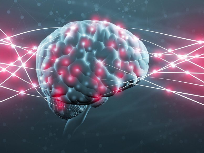
Physically eliminating undesirable or bad memories by changing synapses in the brain might one day be possible.
All memory storage devices, from your brain to the RAM in your computer, store info by altering their physical qualities. Over 130 years earlier, pioneering neuroscientist Santiago Ramón y Cajal first recommended that the brain shops details by rearranging the connections, or synapses, in between nerve cells.
Ever since, neuroscientists have attempted to comprehend the physical modifications associated with memory formation. Picturing and mapping synapses is challenging to do. For one, synapses are very little and securely compacted. Theyre roughly 10 billion times smaller than the tiniest object a basic medical MRI can visualize. There are approximately 1 billion synapses in the mouse brains researchers often use to study brain function, and theyre all the exact same opaque to translucent color as the tissue surrounding them.
Synapses make up the very end of the transmitting neuron, the very beginning of the receiving nerve cell, and the small gap between them.
A brand-new imaging method my colleagues and I established, however, has allowed us to map synapses throughout memory formation. We found that the process of forming brand-new memories changes how brain cells are linked to one another. While some locations of the brain produce more connections, others lose them.
By Don Arnold, USC Dornsife College of Letters, Arts and Sciences
January 15, 2022
When we compared the 3D synapse maps prior to and after memory formation, we found that neurons in one brain area, the anterolateral dorsal pallium, established brand-new synapses while neurons primarily in a 2nd area, the anteromedial dorsal pallium, lost synapses. Associative memories tend to be much stronger than other types of memories, such as mindful memories about what you had for lunch the other day. Associative memories induced by classical conditioning, furthermore, are thought to be analogous to traumatic memories that trigger PTSD. Our research study reveals the function that synaptic connections may play in memory, and might discuss why associative memories can last longer and be kept in mind more strongly than other types of memories.
In theory, this indirectly redesigns the synapses of the brain to make the memory less unpleasant.
Mapping new memories in fish
Formerly, researchers focused on recording the electrical signals produced by neurons. While these research studies have validated that neurons change their reaction to specific stimuli after a memory is formed, they couldnt pinpoint what drives those changes.
To study how the brain physically alters when it forms a new memory, we created 3D maps of the synapses of zebrafish before and after memory development. We picked zebrafish as our test topics because they are large enough to have brains that operate like those of people, but transparent and little enough to provide a window into the living brain.
Zebrafish are especially fitting designs for neuroscience research. Credit: Zhuowei Du and Don B. Arnold
To cause a brand-new memory in the fish, we utilized a kind of learning process called classical conditioning. This includes exposing an animal to 2 different kinds of stimuli concurrently: a neutral one that does not provoke a reaction and an undesirable one that the animal attempts to prevent. When these two stimuli are paired together sufficient times, the animal reacts to the neutral stimulus as if it were the unpleasant stimulus, suggesting that it has actually made an associative memory tying these stimuli together.
As an undesirable stimulus, we carefully heated the fishs head with an infrared laser. When the fish snapped its tail, we took that as a sign that it wished to leave. When the fish is then exposed to a neutral stimulus, a light turning on, tail flicking meant that its remembering what happened when it previously encountered the unpleasant stimulus.
Pavlovs canine is the most well-known example of classical conditioning, in which a pet dog salivates in action to a ringing bell since it has formed an associative memory between the bell and food. Credit: Lili Chin/Flickr
To produce the maps, we genetically engineered zebrafish with nerve cells that produce fluorescent proteins that bind to synapses and make them noticeable. We then imaged the synapses with a customized microscopic lense that utilizes a much lower dosage of laser light than standard gadgets that also utilize fluorescence to generate images. Due to the fact that our microscopic lense triggered less damage to the nerve cells, we were able to image the synapses without losing their structure and function.
This image shows neurons in a live fish brain, with the synapses colored in green. Credit: Zhuowei Du and Don B. Arnold
When we compared the 3D synapse maps prior to and after memory development, we discovered that neurons in one brain area, the anterolateral dorsal pallium, developed new synapses while nerve cells predominantly in a 2nd region, the anteromedial dorsal pallium, lost synapses. This meant that brand-new neurons were combining together, while others ruined their connections. Previous experiments have recommended that the dorsal pallium of fish may be analogous to the amygdala of mammals, where fear memories are saved.
Surprisingly, changes in the strength of existing connections between nerve cells that occurred with memory formation were small and indistinguishable from changes in control fish that did not form new memories. This meant that forming an associative memory includes synapse formation and loss, but not necessarily changes in the strength of existing synapses, as previously believed.
Scientists from Howard Hughes Medical Institute captured video of the firing nerve cells of a child zebrafish as it sees things and tries to move.
Could removing synapses eliminate memories?
Our brand-new technique of observing brain cell function could open the door not just to a much deeper understanding of how memory actually works, however also to potential opportunities for treatment of neuropsychiatric conditions like PTSD and dependency.
Associative memories tend to be much stronger than other types of memories, such as mindful memories about what you had for lunch the other day. Our research study reveals the function that synaptic connections may play in memory, and could describe why associative memories can last longer and be kept in mind more vividly than other types of memories.
In theory, this indirectly redesigns the synapses of the brain to make the memory less painful. This recommends that the underlying memory causing the traumatic response has actually not been removed.
Conceptually connected to classical conditioning, prolonged direct exposure therapy is one way to treat PTSD.
Its still unknown whether synapse generation and loss really drive memory development. My laboratory has actually established innovation that can quickly and precisely get rid of synapses without destructive neurons. We prepare to utilize similar techniques to remove synapses in zebrafish or mice to see whether this changes associative memories.
It might be possible to physically eliminate the associative memories that underlie devastating conditions like PTSD and dependency with these techniques. Prior to such a treatment can even be pondered, however, the synaptic changes encoding associative memories require to be more exactly specified. And there are clearly major ethical and technical difficulties that would require to be resolved. Nevertheless, its tempting to imagine a long run in which synaptic surgical treatment might remove bad memories.
Written by Don Arnold, Professor of Biological Sciences and Biomedical Engineering, USC Dornsife College of Letters, Arts and Sciences.
This post was very first published in The Conversation.


