
Cell sorting through flow cytometry is a technique that allows researchers to determine and sort individual cells based on specific attributes of each cell in order to study them in more detail, evaluate how each cell may respond to a new drug or perform other single cell experiments. Traditionally, cell sorters operate through identification and quantification of specific biomarkers (e.g. proteins) on or within a cell. The brand-new development from BD, understood as BD CellView ™ Image Technology, can record numerous images of private cells flowing through the system at a speed of 15,000 cells per 2nd and also adds a formerly difficult capability of sorting cells based on detailed tiny image analysis of specific cells at this speed.
Image-enabled cell sorting identifies and separates cells of interest out of a complicated swimming pool of cells at high speed. Credit: Tobias Wüstefeld
Innovation breakthrough has possible to transform large range of disciplines from immunology and genomics research to cell-based rehabs.
BD (Becton, Dickinson and Company) (NYSE: BDX), a leading worldwide medical technology business, today revealed that a study performed in partnership with the European Molecular Biology Laboratory (EMBL) and released as the cover story of the January 21st concern of the journal, Science, profiles a brand-new BD innovation in circulation cytometry that includes fluorescence imaging and image-based decisioning to sort individual cells at extremely high speed, based on the visual information of each cell and not exclusively on the type or quantity of biomarkers that exist. The brand-new innovation has the potential to transform immunology, cell biology and genomics research study and make it possible for brand-new cell-based therapeutic discovery.
Cell sorting through circulation cytometry is a technique that allows researchers to recognize and sort individual cells based on particular attributes of each cell in order to study them in more information, assess how each cell may react to a new drug or perform other single cell experiments. Typically, cell sorters operate through identification and quantification of specific biomarkers (e.g. proteins) on or within a cell. The brand-new development from BD, called BD CellView ™ Image Technology, can record numerous pictures of individual cells streaming through the system at a speed of 15,000 cells per 2nd and also includes a previously difficult ability of sorting cells based upon comprehensive tiny image analysis of individual cells at this speed.
By including imaging to the traditional biomarker recognition and metrology, the brand-new innovation not just identifies if and how much of a biomarker is present in the cell, however also its place or how it is distributed within the cell. By imaging the circulation of biomarkers with this innovation, researchers acquire comprehensive info about cells that was formerly undetectable in conventional flow cytometry experiments, which allows them to address complex biological questions, such as how cells grow, engage and operate, or to study specific places of viruses or proteins within a cell, all at an extremely accelerated rate.
“This development essentially relates to a scientist looking into a microscope, determining specific characteristics of a cell of interest, and based on what they see, arranging each private cell for additional analysis– all at a rate of almost 1 million cells every minute. The innovation can examine more than 1,000 times the quantity of data compared to standard flow cytometry approaches, and sort cells at a rate of 15,000 per second based on their images.
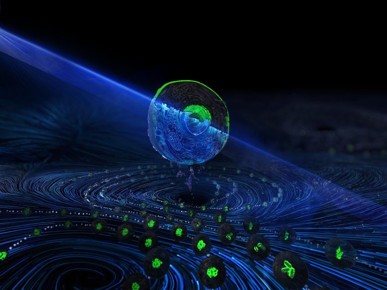
Schematic of image-enabled cell sorting, developed at BD Biosciences and roadway evaluated by EMBL. Credit: BD Biosciences
The new innovation fills an enduring space in biomedical research study by making it possible for scientists to more rapidly view and separate cells with particular, observable traits of interest, which can speed up discovery research and unlock prospective treatments or treatments for illness in a broad variety of fields such as virology and oncology.
” This innovation represents the culmination of more than a decades worth of work from an extremely multidisciplinary group of optical, mechanical, electrical, biomedical and software application engineers and researchers that intended to offer researchers a separated and flexible ability for examining single cells,” said Eric Diebold, worldwide vice president of R&D for BD Biosciences and co-corresponding author of the paper. “We have actually just scratched the surface of the kinds of science that will be allowed with this new high-throughput image-based cell sorting innovation, and we anticipate how BD and the clinical community at-large will leverage it to advance both basic research and the advancement of therapeutics.”
Details of the Study
In the study published in Science, scientists utilized BD CellView ™ Image Technology to study regulators of the NF-? B (nuclear aspect kappa light chain enhancer of triggered B cells) pathway, a protein complex that plays a crucial function in cellular immunity and stress response. The EMBL group measured the activity in this path by tracking the location of RelA, a protein that moves from the cytoplasm into the nucleus of the cell upon activation. Using BD CellView ™ Image Technology, the screen permitted them to determine several novel regulators of this crucial cellular path in a matter of hours, instead of days as would be needed using traditional approaches. This outcome has broad implications for speeding up the speed of genomic research and therapeutic discovery.
” For years, researchers have preferred a system for cell sorting that would enable them to get a detailed photo of a cells inner functions and to separate those with microscopic phenotypes of interest,” stated Dr. Lars Steinmetz, senior scientist at EMBL, teacher of genetics at Stanford University and co-corresponding author of the paper. “This is what BD CellView ™ Image Technology achieves, specifying a new requirement in cell isolation and characterization.
Reference: “Completing a genome-wide screen in less than 9 hours” 21 January 2022, Science.DOI: 10.1126/ science.abj3013.
“This development basically relates to a scientist looking into a microscope, recognizing specific characteristics of a cell of interest, and based on what they see, sorting each private cell for additional analysis– all at a rate of almost 1 million cells every minute.” For years, scientists have actually wanted a system for cell sorting that would allow them to get a comprehensive image of a cells inner workings and to isolate those with microscopic phenotypes of interest,” stated Dr. Lars Steinmetz, senior scientist at EMBL, professor of genetics at Stanford University and co-corresponding author of the paper.