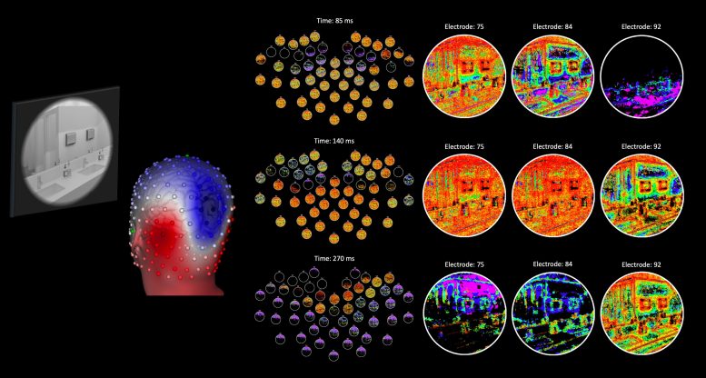
On the right-hand side, each column reveals a close up of the spatiotemporal advancement of the visual code for different electrodes (each row corresponds to a various point in time in milliseconds). Each color represents one of 7 various neural population responses that were mapped to each image area, consequently revealing which neural population best-coded image areas at different points in time. The visual code that is utilized to guide behavior is not stable like an image however rather develops over time with different populations of nerve cells contributing to the code at various points in time. Due to the fact that EEG can determine neural signals at different scalp places, DETI mapping produces a multiplexed view of how different populations of neurons code image features at different locations in images over time– something that was once believed impossible to do with EEG data.
One of the most striking results reported by Hansen and associates is that the brain appears to scan images in a way that stresses different image regions with various neural populations at various points in time.
The research study “Dynamic Electrode-to-Image (DETI) mapping reveals the human brains spatiotemporal code of visual details” has been published in the journal PLOS Computational Biology.
” When seeing any environment, our brains code visual details throughout a large population of nerve cells in a method that allows a range of smart habits. Nevertheless, the visual code that is utilized to direct behavior is not constant like a picture however instead evolves gradually with various populations of nerve cells contributing to the code at various moments. Our DETI mapping method provides a first look into that time-varying code at every place in images,” stated Hansen.
Current advances in voxel-wise encoding analyses based on functional magnetic resonance imaging (fMRI) have actually made it possible for compelling restorations of images based on brain information, however are only able to render a single picture in time due to fMRIs minimal temporal resolution. The DETI mapping treatment presented by Hansen and associates is based upon EEG signals, which manage an opportunity to map the neural code of images with millisecond precision.
To successfully map the visual code to images with EEG data, Hansen and colleagues had to overcome a number of methodological obstacles. Because EEG can measure neural signals at various scalp areas, DETI mapping produces a multiplexed view of how different populations of nerve cells code image functions at various locations in images over time– something that was as soon as believed impossible to do with EEG data.
The mapping information produced by the DETI treatment use new and crucial insights into how the neural code of images progresses gradually. Among the most striking outcomes reported by Hansen and associates is that the brain appears to scan images in such a way that emphasizes different image areas with different neural populations at various points in time. “Such a scanning treatment most likely aids in an early prioritization of the ground plane to support judgments for navigation, with a later emphasis concentrated on landmark organization.”
These findings cause new and interesting concerns connected to how the progressing neural code informs higher level cognitive processes when individuals are engaged in different tasks. “We know that the code for visual details is distributed across a big population of nerve cells, however how that code is dispersed depends on the goals of a given task. What this means is that the brain does not simply create a psychological image based specifically on the environment, but rather creates a representation that best matches the behavioral objectives of the person.” Luckily, DETI mapping allows chances to check out the neural dynamics of task-based visual codes and how those codes ultimately support task-based decision-making.
Referral: “Dynamic Electrode-to-Image (DETI) mapping exposes the human brains spatiotemporal code of visual info” by Bruce C. Hansen, Michelle R. Greene and David J. Field, 27 September 2021, PLoS Computational Biology.DOI: 10.1371/ journal.pcbi.1009456.
Funding: James S. McDonnell Foundation grant, National Science Foundation grant.
On the right-hand side, each column reveals a close up of the spatiotemporal development of the visual code for various electrodes (each row corresponds to a various point in time in milliseconds). Each color represents one of seven various neural population actions that were mapped to each image area, therefore revealing which neural population best-coded image areas at various points in time.
Humans are stepping ever closer to understanding how the brain codes visual details, as scientists have actually now established an approach that maps time-varying brain responses to images to reveal how the brain processes visual info.
Colgate University Neuroscience Professor Bruce C. Hansen worked together with Michelle R. Greene (Bates College), and David J. Field (Cornell University) to introduce vibrant electrode-to-image (DETI) mapping– an analytical method that capitalizes on the high temporal resolution of electroencephalography (EEG) to render maps of visual features that are connected with different neural signals gradually. View a real-time example of neural reactions mapped to an image in the video below.
This video reveals the neural code (at various scalp places) for an example image. The different colors represent responses from various types of neurons. Credit: Bruce Hansen