In the brand-new paper, researchers examined a molecular system potentially accountable for the connection in between Alzheimers illness and circadian rhythms. They measured the activity of immune cells responsible for removing proteins called amyloid-beta that develop as plaques in the brains of people with Alzheimers illness. Using cultures of these cells grown in the lab, they discovered that the immune cells eliminate the amyloid-beta on an oscillating everyday cycle controlled by circadian rhythms. When cells lost that rhythm, the daily cycle vanished. They even more established that the underlying reason for this oscillation was changes in the number of particles of a specific protein, heparan, on the cells surface. The protein they identified responds to body clocks and formerly had actually been shown to play a function in clearing amyloid-beta proteins.
The new findings reveal a system that connects the disturbance of body clocks to Alzheimers disease. The study even more highlights the role of immune cells in this relationship. While more studies will be needed, the brand-new findings present the possibility that, if the everyday clearance of amyloid-beta proteins through this system can be maintained, clients may be less likely to develop Alzheimers disease and to show less severe signs.
Hurley includes, “Understanding how our circadian rhythms can manage cell-surface heparan levels to control the build-up of amyloid-beta may lead to the development of chronotherapeutics that minimize the symptoms of Alzheimers Disease along with other inflammatory diseases.”.
Recommendation: “Circadian control of heparan sulfate levels times phagocytosis of amyloid beta aggregates” by Clark GT, Yu Y, Urban CA, Fu G, Wang C, Zhang F, et al., 10 February 2022, PLoS Genetics.DOI: 10.1371/ journal.pgen.1009994.
Financing: This work was supported by an NIH-National Institute of Biomedical Imaging and Bioengineering Grant U01EB022546 (to J.M.H), an NIH-National Institute of General Medical Sciences Grant R35GM128687 (to J.M.H.), an National Science Foundation CAREER Award 2045674 (to J.M.H.), National Institutes of Health grants 1RF1AG069039 (to C.W.), DK111958 and CA231074 (to R.J.L.), Rensselaer Polytechnic Startup funds (to J.M.H.), a present from the Warren Alpert Foundation (to J.M.H.), and a NIH-National Institute of Aging T32 Fellowship AG057464 (to G.T.C.). The funders had no function in study style, information collection and analysis, choice to publish, or preparation of the manuscript.
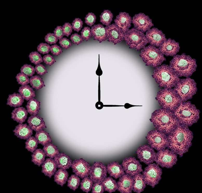
Image Caption: The image of a clock with brightfield macrophage images around it reveals how circadian modifications of cell surface area heparan sulfate proteoglycans, revealed in pink, restrains phagocytosis of fluorescently identified amyloid-beta, shown in green. This image was made using our fluorescent microscopy cell images with a creative making of the heparan sulfate proteoglycans from the app, Wombo.
The findings offer a system that connects Alzheimers disease with circadian rhythm interruptions.
Scientist report that the immune cells responsible for removing a key protein that develops in the brains of clients with Alzheimers disease operate according to everyday body clocks. The discovery, reported by Jennifer Hurley of Rensselaer Polytechnic Institute and colleagues in a brand-new study publishing today (February 10th, 2022) in the journal PLOS Genetics, offers a potential description for the link between Alzheimers illness and disturbances to a persons sleep cycle.
Alzheimers disease is known to be related to interruptions in circadian rhythms, the 24-hour cycle that controls lots of aspects of human behavior and physiology. Sleep interruptions begin years prior to signs of Alzheimers illness appear and are connected to more extreme symptoms and a higher risk of developing the illness.
Image Caption: The image of a clock with brightfield macrophage images around it shows how circadian modifications of cell surface area heparan sulfate proteoglycans, revealed in pink, hampers phagocytosis of fluorescently identified amyloid-beta, shown in green. As time goes on, and we have a decrease in heparan sulfate proteoglycans, we see an increase in phagocytosis, shown by the brilliant green cells revealed on the left of the clock. This image was made using our fluorescent microscopy cell images with an artistic rendering of the heparan sulfate proteoglycans from the app, Wombo. Using cultures of these cells grown in the laboratory, they found that the immune cells clear away the amyloid-beta on an oscillating day-to-day cycle managed by circadian rhythms.