Johns Hopkins Medicine scientists have developed and evaluated a new imaging technique they say will accelerate imaging-based research in the laboratory by enabling investigators to capture images of blood vessels at various spatial scales. Checked in mouse tissues, the method, dubbed “VascuViz,” consists of a quick-setting polymer mixture to fill blood vessels and make them noticeable in numerous imaging methods. Scientists use many different imaging approaches, such as MRI, CT and microscopy to study the role of blood vessels in the laboratory. Incorporating the information readily available in these images has remained a challenge since agents used to make a blood vessel noticeable to one imaging approach can make it invisible on other tools.
Scientists use numerous various imaging techniques, such as MRI, CT and microscopy to study the role of blood vessels in the lab. These images are beneficial for comprehending the dynamics of how tissues develop illness or react to treatment. Integrating the data available in these images has actually stayed an obstacle since representatives utilized to make a blood vessel noticeable to one imaging approach can make it undetectable on other tools. This limits the quantity of information scientists can collect from a single sample.
VascuViz conquers this issue by making the structure of the biggest arteries to the tiniest microvasculature noticeable to a range of imaging tools, which permits researchers to develop a multilayered understanding of blood vessels and associated tissue components with less time and effort.
The advancement of VascuViz is especially helpful in creating computerized visualizations of how complicated biological systems such as the circulatory system work, and is a hallmark of the growing field of “image-based” vascular systems biology.
” Now, rather than utilizing an approximation, we can more specifically estimate features like blood circulation in actual capillary and combine it with complementary info, such as cell density,” says Akanksha Bhargava, Ph.D., postdoctoral fellow in the Pathak Lab within the Department of Radiology and Radiological Science at the Johns Hopkins University School of Medicine. To do this, VascuViz-based measurements are participated in computer system simulations of blood flow, such as the cancer designs Bhargava research studies.
To produce VascuViz, Bhargava evaluated numerous mixes of existing imaging representatives and their viability for various imaging approaches. After multiple models, she found that a CT contrast agent called BriteVu and a fluorescently labeled MRI contrast representative called Galbumin-Rhodamine might be integrated to develop a compound that makes the macro- and microvasculature simultaneously visible when imaging with MRI, CT and optical imaging methods without interference.
With the substance working in test tubes, the scientists then tested it in a variety of mouse tissues, perfusing it through the vascular system of breast cancer models, leg muscles, the brain and kidney tissues. The resulting images of the tissues obtained with MRI, CT and optical microscopy were then combined to produce stunning 3D visualizations of the vasculature and associated elements making up these illness design and organ systems.
Due to VascuVizs affordability and commercially readily available elements, Pathak and his team hope it is worldwide adopted by scientists to help shed brand-new light on different illness involving the vasculature.
Recommendation: “VascuViz: a multimodality and multiscale imaging and visualization pipeline for vascular systems biology” by Akanksha Bhargava, Benjamin Monteagudo, Priyanka Kushwaha, Janaka Senarathna, Yunke Ren, Ryan C. Riddle, Manisha Aggarwal and Arvind P. Pathak, 10 February 2022, Nature Methods.DOI: 10.1038/ s41592-021-01363-5.
Other scientists involved in this study consist of Benjamin Monteagudo, Priyanka Kushwaha, Janaka Senarathna, Yunke Ren, Ryan Riddle and Manisha Aggarwal of the Johns Hopkins University School of Medicine.
This work was supported by the National Cancer Institute (51R01CA196701-05, 1R01CA237597-01A1), the National Institute of Dental & & Craniofacial Research (5R01DE027957-02) and NIH Instrumentation grant (S10OD012287) and the Sidney Kimmel Comprehensive Cancer Center, Quantitative Sciences Pilot Project Grant.
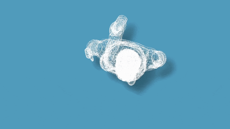
A still from the visual presentation of the result of the VascuViz imaging pipeline. Credit: Johns Hopkins Medicine
Johns Hopkins Medicine scientists have established and tested a brand-new imaging approach they say will speed up imaging-based research study in the lab by permitting detectives to record images of blood vessels at different spatial scales. Checked in mouse tissues, the approach, called “VascuViz,” consists of a quick-setting polymer mixture to fill blood vessels and make them noticeable in several imaging techniques.
The report was released on February 10, 2022, in Nature Methods.
” Usually, if you desire to collect information on blood vessels in a provided tissue and combine it with all of its surrounding context like the structure and the types of cells growing there, you have to re-label the tissue numerous times, get several images and piece together the complementary information,” states Arvind Pathak, Ph.D., teacher of radiology, biomedical and electrical engineering and member of the Sidney Kimmel Comprehensive Cancer at the Johns Hopkins University School of Medicine. “This can be a lengthy and costly procedure that risks destroying the tissues architecture, precluding our ability to use the combined information in novel methods.”