Utilizing a new technique, researchers have discovered that cells lose about 4% of their mass as they get in cellular division. They are essentially taking out the garbage to give their offspring a fresh start.
Cells may utilize this strategy to clear out poisonous byproducts and provide their offspring a fresh start.
MIT researchers have actually found that before cells begin to divide, they do a little cleanup, tossing out particles that they appear not to need anymore.
Using a brand-new technique they established for determining the dry mass of cells, the scientists discovered that cells lose about 4 percent of their mass as they enter cellular division. The scientists believe that this emptying of trash assists cells to give their offspring a “fresh start,” devoid of the moms and dad cells accumulated junk.
About 10 years ago, they had found that they could calculate a cells dry mass if they first measured the cell in typical water and then in heavy water (which consists of deuterium instead of regular hydrogen). In cells going through mitosis, the scientists utilized their new strategy to study what takes place to cell mass and structure throughout that process. Other research studies that utilized quantitative phase microscopy suggested that cells might maintain or lose dry mass early in cell department.
To their surprise, the scientists discovered that the dry mass of cells in fact decreases when they go into the cell division cycle. The scientists also found that the density of the dry mass increases as the cells lose dry mass, leading them to believe that the cells are losing low-density particles such as lipoproteins or lipids.
” Our hypothesis is that cells might be tossing out things that are developing up, harmful components or just things that dont operate effectively that you dont desire to have there. It could allow the newborn cells to be born with more practical contents,” says Teemu Miettinen, an MIT research researcher and the lead author of the new study.
Scott Manalis, the David H. Koch Professor of Engineering in the departments of Biological Engineering and Mechanical Engineering, and a member of the Koch Institute for Integrative Cancer Research, is the senior author of the paper, which was published on May 10, 2022, in the journal eLife. MIT biological engineering undergraduates Kevin Ly and Alice Lam are also authors of the paper.
Measuring mass
Measuring the dry mass of a cell– the weight of its contents not including the water– is typically done using a microscopy method called quantitative phase microscopy. This method can determine cell development, however it does not reveal info about the molecular content of the dry mass and it is difficult to utilize with cells that grow in suspension.
Manalis laboratory has actually previously developed a strategy for measuring the buoyant mass of cells, which is their mass as they drift in a fluid such as water. This technique measures buoyant mass by flowing cells through a channel embedded in a vibrating cantilever, which can be done repeatedly to track changes in a particular cells mass over lots of hours or days.
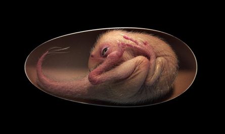
MIT researchers have found that before cells start to divide, they toss waste items. In this image, the magenta represents DNA, and the green represents a lysosomal marker on the surface area of the cells, which is a sign of lysosomal exocytosis. Credit: Courtesy of the researchers
For their new study, the scientists desired to adapt the strategy so that it could be used to determine the dry mass of cells, in addition to the density of the dry mass. About 10 years ago, they had actually found that they could calculate a cells dry mass if they initially determined the cell in regular water and after that in heavy water (which includes deuterium rather of regular hydrogen). These two measurements can be used to determine the cells dry mass.
Nevertheless, heavy water is harmful to cells, so they were just able to get a single measurement per cell. Last year, Miettinen set out to see if he might develop a system in which cells could be determined consistently with minimal direct exposure to heavy water.
In the system he created, cells are exposed to heavy water extremely quickly as they stream through microfluidic channels. It takes just one 2nd for a cell to completely exchange its water material, so the researchers might measure the cells mass when it was complete of heavy water, compare it to the mass in normal water, and after that compute the dry mass.
” Our idea was that if we minimize the cells exposure to the heavy water, we could craft the system so that we might repeat this measurement over prolonged time durations without hurting the cell,” Miettinen states. “That allowed us for the very first time to track not simply the dry mass of a cell, which is what others do using tiny approaches, however likewise the density of the dry mass, which notifies us of the cells biomolecular structure.”
The scientists showed that their dry mass measurements qualitatively agreed with previous work using quantitative phase microscopy. And, in addition to supplying density of the dry mass, the MIT groups method makes it possible for greater temporal resolution, which showed to be beneficial for exposing dynamics during mitosis (cell department).
Getting the garbage
In cells undergoing mitosis, the researchers used their new technique to study what takes place to cell mass and structure throughout that procedure. In a 2019 paper, Miettinen and Manalis found that buoyant mass increases somewhat as mitosis begins. Other research studies that used quantitative stage microscopy recommended that cells might maintain or lose dry mass early in cell department.
In the brand-new research study, the MIT team measured three kinds of cancer cells, which are easier to study because they divide more regularly than healthy cells. To their surprise, the scientists discovered that the dry mass of cells really reduces when they go into the cell department cycle. This mass is regained later, prior to department is total.
More experiments exposed that as cells go into mitosis, they ramp up activity of a procedure called lysosomal exocytosis. Lysosomes are cell organelles that break down or recycle cellular waste products, and exocytosis is the process they use to reject any particles that arent needed any longer.
The researchers also found that the density of the dry mass increases as the cells lose dry mass, leading them to think that the cells are losing low-density molecules such as lipoproteins or lipids. They assume that cells use this process to clear out harmful particles prior to dividing. “What we are seeing is that cells might be attempting to throw out damaged parts prior to dividing,” Miettinen states.
The scientists hypothesize that their findings may assist describe why neurons, which do not divide, are most likely to accumulate toxic proteins such as Tau or amyloid beta, which are linked to the advancement of Alzheimers disease.
The findings could also pertain to cancer: Cancer cells can expel some chemotherapy substance abuse exocytosis, assisting them to end up being resistant to the drugs. In theory, preventing exocytosis from happening prior to cellular division could assist to make cancer cells more vulnerable to such drugs.
” There are diseases where we might desire upregulate exocytosis, for instance in neurodegenerative diseases, however then there are diseases like cancer where maybe we desire to dial it down,” Miettinen states. “In the future, if we could better understand the molecular system behind this, and discover a method to activate it beyond mitosis or prevent it throughout mitosis, we could really have a new toggle to utilize when treating disease.”
Recommendation: “Single-cell monitoring of dry mass and dry mass density exposes exocytosis of cellular dry contents in mitosis” by Teemu P Miettinen, Kevin S Ly, Alice Lam, Scott R Manalis, 10 May 2022, eLife.DOI: 10.7554/ eLife.76664.
The research was funded by the MIT Center for Cancer Precision Medicine, the Virginia and D.K. Ludwig Fund for Cancer Research, the Cancer Systems Biology Consortium, and the Koch Institute Support (core) Grant from the National Cancer Institute.