Various imaging approaches are used to catch info about tissue, cells, and subcellular structures. A single technique can just record information about either the structure or function of the tissue and looking in detail at a nanometer scale (a nanometer is one billionth of a meter) means researchers lose details about the larger surroundings. This suggests that to get an overall understanding of the tissue, imaging techniques require to be integrated.
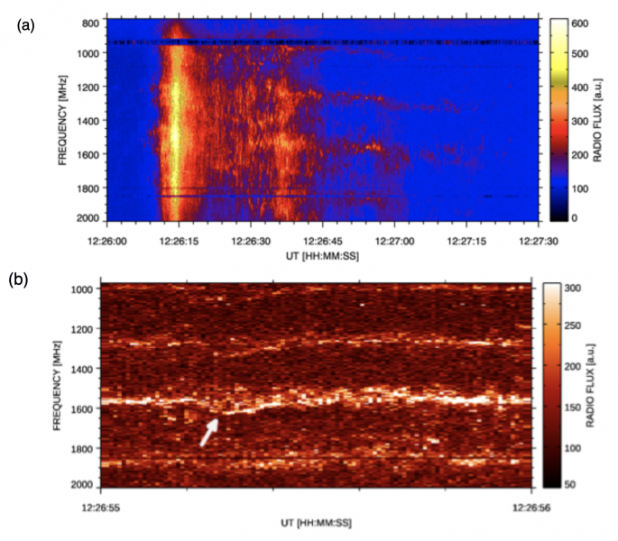
After the mice were euthanized, brain tissue samples were imaged using different methods consisting of synchrotron X-ray tomography, which records samples up to a number of millimeters in length.
Numerous imaging methods are used to catch details about tissue, cells, and subcellular structures. A single technique can just catch details about either the structure or function of the tissue and looking in information at a nanometer scale (a nanometer is one billionth of a meter) suggests researchers lose details about the wider environments. This suggests that to get a total understanding of the tissue, imaging techniques require to be integrated.
In their study, the researchers established a method that integrates 7 imaging approaches, including in vivo imaging, synchrotron X-ray, and volume electron microscopy. They demonstrated their technique by imaging two different areas of the brain in mice– the olfactory bulb and the hippocampus.
Importantly, the technique could be applied to other locations of the brain or parts of the body, offering researchers with a more comprehensive understanding of numerous various biological structures and tissues.
Each action of the imaging procedure supplies various details. To start with, the researchers utilized in vivo calcium imaging to imagine nerve cells in specific areas of the brain and see which neurons were active when the mice were exposed to odors.
Four pictures of the same area of the mouse olfactory bulb glomerular layer, using various imaging approaches. Credit: Carles Bosch
After the mice were euthanized, brain tissue samples were imaged utilizing different techniques consisting of synchrotron X-ray tomography, which catches samples approximately a number of millimeters in length. This scale is sufficient for scientists to see whole neural networks and likewise where other structures or particular cells sit within the larger context of the sample. Notably, this method does not harm the sample so it can be imaged again using another technique.
The scientists then selected locations of particular interest to be imaged with electron microscopy, catching detailed detail at a high resolution. At some target areas, this might map details as small as 10 nanometers, enabling researchers to see tiny structures like the specific synapses that link nerve cells.
Using computer system algorithms, they combined the results to create a complete map of the structure and function of the area of the brain they were studying, up to a couple of cubic millimeters.
Carles Bosch, first author and Principal Laboratory Research Scientist in the Sensory Circuits and Neurotechnology Laboratory at the Crick states: “Our technique supplies a dependable method to overcome the obstacle of imaging structures at greatly different scales. Our company believe it will be a powerful tool to investigate neuronal circuits in the brains of mammals along with the structure and function of other tissues.”
Andreas Schaefer, senior author and head of the Sensory Circuits and Neurotechnology Laboratory at the Crick states: “Were interested in applying this method to the brain, where it is necessary to collect information about entire neural networks which are several millimeters in length alongside details about particular neurons and synapses.
” But it also has excellent prospective to be useful in other settings, like cancer biology where scientists intend to understand the activity of particular cells in the context of the wider tumor.”
The scientists partnered with the electron microscopy team at the Crick, and also worked with synchrotron X-ray teams at the Diamond Light Source in Oxfordshire, The European Synchrotron in France and the Paul Scherrer Institut in Switzerland.
The team will continue this research study, using this imaging approach to reveal more detail about the olfactory bulb in addition to working to more improve the strategy.
Referral: “Functional and multiscale 3D structural examination of brain tissue through correlative in vivo physiology, synchrotron micro-tomography and volume electron microscopy” by Bosch, C. et al., 25 May 2022, Nature Communications.DOI: 10.1038/ s41467-022-30199-6.
Scientists at the Francis Crick Institute have established an imaging method to record info about the structure and function of brain tissue at subcellular level– a few billionths of a meter, while likewise catching details about the surrounding environment.
The unique technique detailed in the journal Nature Communications today (May 25, 2022), conquers the difficulties of imaging tissues at various scales, allowing researchers to see the surrounding cells and how they work, so they can develop a complete image of neural networks in the brain.