DVP links data-rich imaging of cell culture or archived patient biobank tissues with deep learning-based cell division and machine learning-based identification of cell types and states. Subsequent bioinformatics data analysis allows information mining to discover protein signatures providing molecular insights into proteome variation in health and illness states at the level of single cells. It exposes a cosmos of particles inside these cancer cells,” states Andreas Mund, Associate Professor at the Novo Nordisk Foundation Center for Protein Research (CPR) and part of Professor Matthias Manns group that led this advancement at CPR and the Max Planck Institute for Biochemistry.
It enables researchers to examine interactions in between cancer cells and their surrounding cells with significant ramifications for future clinical cancer treatment. Simply the normal or diseased cells of a particular type are analyzed by mass spectroscopy, mapping the protein landscape, and understanding the systems of health and disease.
By University of Copenhagen – The Faculty of Health and Medical Sciences
June 27, 2022
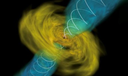
Deep Visual Proteomics principle and workflow Clockwise: Deep Visual Proteomics (DVP) combines high-resolution imaging, expert system (AI)- assisted image analysis for single-cell category and isolation with a novel ultra-sensitive proteomics workflow. DVP links data-rich imaging of cell culture or archived patient biobank tissues with deep learning-based cell division and maker learning-based identification of cell types and states. (Un) supervised AI-classified cellular or subcellular items of interest go through automated laser microdissection and mass spectrometry (MS)- based proteomic profiling. Subsequent bioinformatics data analysis makes it possible for information mining to discover protein signatures offering molecular insights into proteome variation in health and illness states at the level of single cells. Credit: MPI of Biochemistry
How do some patients establish resistance to cancer treatment? The brand-new technique understood as “Deep Visual Proteomics” might have the ability to help doctors get closer to a response and recognize cancer growth vulnerabilities.
It is never simple for physicians to determine why particular illnesses develop in our bodies. Old age, risky habits such as smoking, and genetics can all play a role.
The specific, particular causes of severe diseases such as cancer remain unknown.
Now, a cutting-edge approach called “Deep Visual Proteomics” might be able to assist alter that. A global group of researchers led by Copenhagen University created the technique, which was just recently applied to cancer cells in a new study released in the top clinical journal Nature Biotechnology.
” Our brand-new concept, Deep Visual Proteomics, might end up being a game-changer for molecular pathology in the health centers. With this technique, we can determine countless proteins and identify how many of them are there,” describes Andreas Mund, very first author of the new study.
” We do this by taking a tissue sample and analyzing just the growth cells in it. This list of proteins is called proteome. These proteomes expose the systems that drive growth advancement and straight expose new healing targets from a single tissue slice of a cancer client biopsy. It exposes an universe of particles inside these cancer cells,” states Andreas Mund, Associate Professor at the Novo Nordisk Foundation Center for Protein Research (CPR) and part of Professor Matthias Manns team that spearheaded this advancement at CPR and limit Planck Institute for Biochemistry.
Essential to pathology departments
The reason that the scientists are so interested in proteins is that they actually are a few of the most important pieces of the puzzle for almost all diseases. Proteins are frequently referred to as the workhorses of the cell.
” When something goes wrong inside our cells and we become sick, you can be sure that proteins are associated with a wide variety of different methods. Due to the fact that of this, mapping the protein landscape can help us figure out why a growth could establish in a specific client, what vulnerabilities that tumor has, and likewise what treatment method might prove the most beneficial,” states Matthias Mann, teacher.
In the new study, the researchers used “Deep Visual Proteomics” to cells from patients with acinic cell cancer and cancer malignancy. This was performed in cooperation with scientists at the Zealand University Hospital, Roskilde.
” This special technique combines tissue architecture with the expression of countless proteins particular for chosen cells. It makes it possible for researchers to investigate interactions in between cancer cells and their surrounding cells with major implications for future scientific cancer treatment. Just recently, we detected an extremely complicated scientific case with 2 different components and the arise from DVP analysis,” states Lise Mette Rahbek Gjerdrum, consultant and scientific research study associate professor at Department of Pathology, Zealand University Hospital and Department of Clinical Medicine, University of Copenhagen.
Digital pathology, deep knowing, microscopy, and mass spectrometry
Afterward, device learning algorithms are utilized to categorize cells accurately before laser microdissections and single-cell collection. Just the unhealthy or normal cells of a specific type are evaluated by mass spectroscopy, mapping the protein landscape, and comprehending the systems of health and disease.
” Using this technology, we can efficiently connect the physiological qualities of cells seen under microscopes with the functions of proteins. This was not previously possible and we are extremely persuaded that this technique can be applied to other diseases, not simply cancer,” says Andreas Mund.
Reference: “Deep Visual Proteomics defines single-cell identity and heterogeneity” by Andreas Mund, Fabian Coscia, András Kriston, Réka Hollandi, Ferenc Kovács, Andreas-David Brunner, Ede Migh, Lisa Schweizer, Alberto Santos, Michael Bzorek, Soraya Naimy, Lise Mette Rahbek-Gjerdrum, Beatrice Dyring-Andersen, Jutta Bulkescher, Claudia Lukas, Mark Adam Eckert, Ernst Lengyel, Christian Gnann, Emma Lundberg, Peter Horvath and Matthias Mann, 19 May 2022, Nature Biotechnology.DOI: 10.1038/ s41587-022-01302-5.