Unusual illness of the retina are frequently triggered by inherited anomalies in genes revealed by PRCs. These private illness are rare, together they are the leading cause of loss of sight in working-age grownups.
Some of these genetic variants were understood to be connected to eye illness, but remarkably, a number of relatively common genetic variants were near genes understood to trigger uncommon genetic eye diseases when interrupted. In one case, the researchers were able to check out how combinations of common variants near genes known to be included in unusual eye illness change the structure of the retina. This gives more self-confidence when looking into specific uncommon illness collections to see how these specific common variations may impact illness.
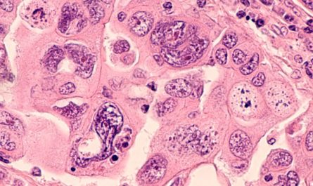
Large-scale information allows brand-new insights into unusual eye conditions. Credit: Karen Arnott/EMBL-EBI
Scientists at EMBL-EBI delve into the UK Biobank information to reveal brand-new information about unusual diseases of the eye.
Scientists have actually performed a thorough evaluation of image and genomic information from the UK Biobank to gain a deeper understanding of uncommon eye diseases. Among these diseases are retinal dystrophies, which is a collective term for genetic conditions that impact the retina and likewise happen to be the main reason for blindness among working-age grownups.
The retina is located at the back of the eye and is a complicated tissue composed of several layers. It operates by receiving light and changing it into a signal that can be understood by the brain. Each layer of the retina is consisted of different cell types, each playing a distinct function in the conversion of light.
For this research study released in the journal PLOS Genetics, the scientists focused on photoreceptor cells (PRCs), which are light-detecting cells found in the retina. These cells can be non-invasively imaged using optical coherence tomography (OCT), a service now commonly offered in many opticians. Utilizing OCT image information and genomic data saved in the UK Biobank, researchers were able to produce the biggest genome-wide association study of PRCs.
Rare retinal dystrophies
Rare illness of the retina are often triggered by acquired mutations in genes revealed by PRCs. These anomalies cause the retina to work improperly, resulting in sight disability and even blindness. Although these private diseases are rare, together they are the leading cause of loss of sight in working-age adults.
” We had access to paired images and genotype data at a scale that had actually not been seen in a study of this kind,” stated Hannah Currant, previous Ph.D. student at EMBLs European Bioinformatics Institute (EMBL-EBI) and Postdoctoral Fellow at the Novo Nordisk Foundation Center for Protein Research (CPR) University of Copenhagen. “Access to this massive quantity of information was vital to the study and enabled us to recognize hereditary links to uncommon retinal dystrophies. This work has actually recognized brand-new avenues for research and created brand-new concerns about rare retinal dystrophies.”
Linking genotype and phenotype
OCT produces high-resolution images that can be used to identify the various layers and structures within the retina. These images are commonly used in the clinic to help the diagnosis of eye disorders. For this study, the researchers utilized OCT images and the matching genomic and medical details of over 30,000 participants saved in the UK Biobank.
” The UK Biobank is an abundant, invaluable resource that has massive capacity to make it possible for genomic medicine,” stated Ewan Birney, Deputy Director General of the European Molecular Biology Laboratory (EMBL). “There is so much potential simply waiting to be launched from the data stored there, which lets us both comprehend human biology and how and when it fails in disease.”
Driving genomic medicine
The researchers carried out genome-wide association research studies (GWAS) on the UK Biobank data to search for hereditary variations linked to differences in the density of the PRC layers. This led them to determine genomic variations associated with the thickness of one or more of the PRC layers, including those with previous associations with recognized eye diseases. The newly recognized genomic associations are saved and can be openly accessed through the GWAS Catalog.
A few of these genetic variants were known to be linked to eye illness, however remarkably, a variety of reasonably common hereditary variations were near genes known to cause uncommon genetic eye illness when disrupted. In one case, the researchers were able to explore how combinations of common versions near genes known to be included in unusual eye diseases change the structure of the retina. When looking into specific rare disease collections to see how these specific common variations might impact disease, this offers more self-confidence.
” Systematic bioinformatic analysis of massive participant information accomplices is driving the future of genomic medication,” said Omar Mahroo, Professor of Retinal Neuroscience at University College London and Consultant Ophthalmologist at Moorfields Eye Hospital. “Having access to these information and having the ability to make these connections between disease phenotypes and hereditary variation will open numerous new chances for contemporary illness medical diagnosis and therapeutics.”
Reference: “Sub-cellular level resolution of common hereditary variation in the photoreceptor layer determines continuum between rare illness and typical variation” by Hannah Currant, Tomas W. Fitzgerald, Praveen J. Patel, Anthony P. Khawaja, UK Biobank Eye and Vision Consortium, Andrew R. Webster, Omar A. Mahroo and Ewan Birney, 27 February 2023, PLOS Genetics.DOI: 10.1371/ journal.pgen.1010587.
The research study was funded by the Wellcome Trust.