Human Brain Project (HBP) scientists used a distinct multi-scale method, including various speculative techniques, to investigate the complex company of the brains connectome. Their findings, linked within the Julich Brain Atlas for referral, expose fresh insights into the connectivity and function of various brain regions.
Microscopy approaches like TPFM provide sub-micrometer resolution images of little brain volumes exposing microstructures of the brains cerebral cortex, but they have their restrictions in disentangling fibers linking distant brain areas, which construct the deep white matter structures. The three-dimensional atlas contains more than 250 cytoarchitectonic maps of brain areas and forms the centerpiece of the HBPs Multilevel Human Brain Atlas. “Our brain atlas allows us to pinpoint exactly where in the brain we find these microstructures,” discusses Amunts.
Human Brain Project (HBP) scientists used a distinct multi-scale technique, integrating different speculative methods, to examine the complex company of the brains connectome. They combined strategies like physiological and diffusion magnetic resonance imaging, two-photon fluorescence microscopy, and 3D Polarized Light Imaging to comprehend and visualize nerve fibers at different spatial scales. Their findings, linked within the Julich Brain Atlas for referral, reveal fresh insights into the connection and function of different brain areas.
Researchers from the Human Brain Project utilized a multi-scale technique, combining various imaging approaches to understand the human brains connectome, or the interconnected structure, from the molecular to the macro level. They utilized 3D Polarized Light Imaging to envision nerve fibers and put their findings within the Julich Brain Atlas to spatially reference the information, exposing new insights about the brains company and working.
To comprehend how our brain works, there is no getting around examining how different brain areas are gotten in touch with each other by nerve fibers. In the journal Science, scientists of the Human Brain Project (HBP) review the present state of the field, provide insights on how the brains connectome is structured on different spatial scales– from the molecular and cellular to the macro level– and assess existing approaches and future requirements for comprehending the connectomes complex company.
” It is insufficient to study brain connectivity with one single technique, or perhaps two,” says author and HBP Scientific Director Katrin Amunts, who leads the Institute of Neuroscience and Medicine (INM-1) at Forschungszentrum Jülich and the C. & & O. Vogt Institute of Brain Research at the University Hospital Düsseldorf. “The connectome is embedded at multiple levels. To understand its structure, we require to look at a number of spatial scales simultaneously by combining different experimental methods in a multi-scale method and by integrating the acquired information into multilevel atlases such as the Julich Brain Atlas that we have actually established.”
Markus Axer from Forschungszentrum Jülich and the Physics Department of the University of Wuppertal, who is the very first author of the Science post, has with his team at INM-1 established a distinct method called 3D Polarized Light Imaging (3D-PLI) to visualize nerve fibers at tiny resolution. The researchers trace the three-dimensional courses of fibers throughout serial brain sections with the aim of establishing a 3D fiber atlas of the whole human brain.
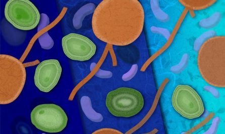
Information of a human brain section revealing the architecture of fibers down to single axons in the hippocampus, exposed by 3D Polarized Light Imaging. Colors represent 3D fiber orientations highlighting pathways of person fibers and systems. Credit: Markus Axer and Katrin Amunts, INM-1, Forschungszentrum Jülich
Together with other HBP scientists from Neurospin in France and the University of Florence in Italy, Axer and his group have recently imaged the exact same tissue block from a human hippocampus utilizing a number of various approaches: physiological and diffusion magnetic resonance imaging (aMRI and dMRI), two-photon fluorescence microscopy (TPFM) and 3D-PLI, respectively.
Microscopy techniques like TPFM supply sub-micrometer resolution images of small brain volumes exposing microstructures of the brains cerebral cortex, however they have their limitations in disentangling fibers linking distant brain regions, which build the deep white matter structures. This is a lot more real for electron microscopic measurements, which make it possible for nanometre-resolved insights into a cubic millimeter of brain tissue. On the other hand, dMRI can be utilized for tractography at the whole-brain level– imagining white matter connections– but can not deal with individual fibers or small systems.
” 3D-PLI functions as a bridge between micro and macro methods,” states Amunts. “This is because 3D-PLI solves the fiber architecture at high resolution and, at the same time, enables imaging of whole-brain sections that we can then reconstruct in 3D to trace fiber connections.”
Integrating 3d-pli, tpfm, and dmri enabled the researchers to superimpose the three techniques within the very same recommendation area. “This integration of data was just made possible by imaging one and the exact same tissue sample,” discusses Axer. The human hippocampus block took a trip from Germany to France, back to Germany, and lastly to Italy, being processed and imaged in different laboratories benefiting from the regional, highly specialized equipment.
The researchers then used the Julich Brain Atlas to spatially anchor their data in an anatomical referral area. The three-dimensional atlas consists of more than 250 cytoarchitectonic maps of brain areas and forms the centerpiece of the HBPs Multilevel Human Brain Atlas. “Our brain atlas allows us to pinpoint exactly where in the brain we discover these microstructures,” explains Amunts. The dataset is openly accessible via the HBPs EBRAINS infrastructure and can be browsed in an interactive atlas audience.
The researchers multi-scale approach combining numerous methods at various spatial scales to unravel the human connectome is unique and provides amazing new insights into how the human brain works.
Although the hippocampus reconstruction is a lighthouse project, there are a number of global efforts continuous (or about to begin) that need to be orchestrated at an open atlas level to make it possible for the combination of multi-scale data. Amunts and Axer stress that this is a prerequisite for revealing the concepts of connection within the experimentally accessible range of scales– from axons to paths. Simply put, an integrated multi-scale approach that integrates micro and macro techniques is needed to describe and understand the embedded organization of the human brain. This requires crucial reassessment of present method, including tractography, the authors say.
Recommendation: “Scale matters: The embedded human connectome” by Markus Axer and Katrin Amunts, 3 November 2022, Science.DOI: 10.1126/ science.abq2599.