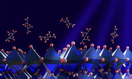
MIT biologists found that a scaffolding protein called TCOF1 is accountable for the development of a biomolecular condensate called the fibrillar center, which forms within the cell nucleolus, represented in pink inside the purple nucleus. Credit: iStock
A single protein can self-assemble to construct the scaffold for a biomolecular condensate that makes up a key nucleolar compartment.
Inside all living cells, loosely formed assemblies called biomolecular condensates perform lots of important functions. It is not well understood how proteins and other biomolecules come together to form these assemblies within cells.
MIT biologists have actually now found that a single scaffolding protein is responsible for the development of among these condensates, which forms within a cell organelle called the nucleolus. Without this protein, called TCOF1, this condensate can not form.
The findings could help to explain a significant evolutionary shift, which took location around 300 million years back, in how the nucleolus is arranged. Until that point, the nucleolus, whose role is to assist develop ribosomes, was divided into two compartments. However, in amniotes (which consist of birds, reptiles, and mammals), the nucleolus established a condensate that acts as a third compartment. Biologists do not yet completely understand why this shift took place.
Evolutionary Changes in the Nucleolus
” If you look across the tree of life, the standard structure and function of the ribosome has stayed quite static; however, the procedure of making it keeps developing. Our hypothesis for why this procedure keeps developing is that it might make it much easier to assemble ribosomes by separating the different biochemical responses,” says Eliezer Calo, an associate teacher of biology at MIT and the senior author of the research study.
Now that the researchers know how this condensate, understood as the fibrillar center, kinds, they might be able to more quickly study its function in cells. The findings likewise offer insight into how other condensates may have originally developed in cells, the scientists say.
Former MIT college student Nima Jaberi-Lashkari PhD 23 and Byron Lee PhD 23 are the lead authors of the paper, which was released just recently in the journal Cell Reports. Former MIT research study associate Fardin Aryan is also an author of the paper.
Condensate Formation
Lots of cell functions are performed by membrane-bound organelles, such as lysosomes and mitochondria, but membraneless condensates likewise perform critical tasks such as gene guideline and stress action. In some cases, these condensates form when needed and after that liquify when they are completed with their job.
” Almost every cellular procedure that is essential for the performance of the cell has been associated in some way with condensate development and activity,” Calo says. “However, its not effectively figured out how these condensates form.”
In a 2022 study, Calo and his colleagues recognized a protein area that seemed to be associated with forming condensates. In that study, the researchers utilized computational techniques to determine and compare stretches of proteins called low-complexity regions (LCRs), from many different species. LCRs are sequences of a single amino acid duplicated sometimes, with a couple of other amino acids sprayed in.
That work also exposed that a nucleolar protein understood as TCOF1 includes many glutamate-rich LCRs that can help scaffold biomolecular assemblies.
In the new research study, the researchers discovered that whenever TCOF1 is revealed in cells, condensates form. These condensates always consist of proteins normally found within a specific condensate called the fibrillar center (FC) of the nucleolus. The FC is known to be included in the production of ribosomal RNA, a crucial element of ribosomes, the cell complex responsible for constructing all cellular proteins.
However, in spite of its importance in assembling ribosomes, the fibrillar center appeared only around 300 million years earlier; single-celled organisms, invertebrates, and the earliest vertebrates (fish) do not have it.
The new research study suggests that TCOF1 was important for this shift from a “bipartite” to “tripartite” nucleolus. The scientists discovered without TCOF1, cells form only 2 nucleolar compartments. Additionally, when the researchers added TCOF1 to zebrafish embryos, which typically have bipartite nucleoli, they could cause the formation of a 3rd compartment.
” More than just producing that condensate, TCOF1 rearranged the nucleolus to acquire tripartite residential or commercial properties, which shows that whatever chemistry that condensate was bringing to the nucleolus was enough to change the composition of the organelle,” Calo says.
Scaffold Evolution
The scientists likewise found that the essential region of TCOF1 that assists it form scaffolds is the glutamate-rich low-complexity regions. These LCRs appear to connect with other glutamate-rich regions of neighboring TCOF1 molecules, assisting the proteins put together into a scaffold that can then draw in other proteins and biomolecules that help form the fibrillar center.
” Whats truly amazing about this work is that it offers us a molecular deal with to manage a condensate, present it into a species that doesnt have it, and likewise get rid of it in a types that does have it. That might actually assist us open the structure-to-function relationship and figure out what is the function of the third compartment,” Jaberi-Lashkari says.
Based upon the findings of this research study, the researchers assume that cellular condensates that emerged earlier in evolutionary history may have originally been scaffolded by a single protein, as TCOF1 scaffolds the fibrillar center, however slowly evolved to become more complex.
” Our hypothesis, which is supported by the data in the paper, is that these condensates might stem from one scaffold protein that behaves as a single element, and in time, they become multicomponent,” Calo states.
Implications for Disease Research
The formation of specific types of biomolecular condensates has also been connected to conditions such as ALS, Huntingtons disease, and cancer.
” In all of these circumstances, what our work presents is this question of why are these assemblies forming, and what is the scaffold in these assemblies? And if we can much better understand that, then I believe we have a better manage on how we could deal with these diseases,” Lee states.
Recommendation: “An evolutionarily nascent architecture underlying the development and emergence of biomolecular condensates” by Nima Jaberi-Lashkari, Byron Lee, Fardin Aryan and Eliezer Calo, 15 August 2023, Cell Reports.DOI: 10.1016/ j.celrep.2023.112955.
The research was moneyed by the National Institutes of Health, the National Institute of General Medical Sciences, the Pew Charitable Trusts, and the National Cancer Institute.
In amniotes (which include reptiles, birds, and mammals), the nucleolus established a condensate that acts as a 3rd compartment. In a 2022 research study, Calo and his coworkers determined a protein area that appeared to be included in forming condensates. In the brand-new study, the scientists found that whenever TCOF1 is expressed in cells, condensates form. These condensates constantly include proteins generally discovered within a specific condensate understood as the fibrillar center (FC) of the nucleolus. The researchers found without TCOF1, cells form only 2 nucleolar compartments.