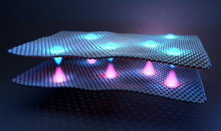
The greyscale pictures represent 2D forecasts of several views of the imaging scaffold attached to a target protein; the color image illustrates the 3D restoration derived from 2D forecasts. An electron microscope image of scaffolds attached to the protein KRAS (background). In cryo-EM, researchers use a cryo-electron microscope to send a beam of electrons through frozen samples of product, leaving behind an image of the thousands of particles– such as proteins– in the sample. Computer system processing reconciles all of those photos to formulate a right 3D image– separating the background, organizing images with similar orientations together, and producing a high-resolution, 3D image of a single molecule.
The scientists checked their scaffold by attempting to develop images of a protein called KRAS which encourages cells to proliferate.
Now, biochemists from the University of California, Los Angeles (UCLA), dealing with pharmaceutical market researchers, have established a service that will make it possible for cryo-EM to acquire high-quality pictures of smaller sized protein molecules, too. The scientists crafted a 20-nanometer, cube-shaped protein structure, called a scaffold, with rigid tripod-like protrusions that hold the small proteins in place.
The scaffold can be digitally eliminated from the photo when the imaging is being processed, leaving a composite 3D picture of just the small protein scientists are evaluating.
An electron microscopic lense image of scaffolds connected to the protein KRAS (background). The left circle highlights one imaging scaffold, the second displays the 3D structure of the imaging scaffold bound to KRAS, and the third shows a close-up of KRAS connected to the cancer drug AMG510. Credit: Roger Castells-Graells/UCLA
Small and medium-sized proteins are a hot point for research on possible brand-new drugs that might one day be utilized to combat some of the most intractable human diseases. The advance, which was evaluated on a protein that researchers are studying for its possible use in cancer treatments, can be tailored for practically any little protein. Researchers anticipate that expanding cryo-EMs imaging abilities will help them determine specific places on proteins that they can target for healing purposes.
A paper about the new research was just recently released in Proceedings of the National Academy of Science (PNAS).
How Cryo-EM Works
In cryo-EM, scientists utilize a cryo-electron microscopic lense to send a beam of electrons through frozen samples of material, leaving an image of the thousands of particles– such as proteins– in the sample. The particles are imaged exactly as they lie in the sample, producing countless 2D photos of the particle drawn from different angles. Computer processing reconciles all of those photos to create a proper 3D image– separating the background, organizing images with comparable orientations together, and creating a high-resolution, 3D image of a single particle.
When it comes to imaging the smallest protein molecules, their tiny size makes it impossible to determine their orientations in the images, which results in relatively low-resolution images.
In previous research studies, scientists tried to solve the issue by connecting little molecules to bigger scaffolds, however those experiments demonstrated that if the little particles were attached too flexibly, they would protrude from the scaffold at different angles and orientations– which would still produce fuzzy images.
” The images are fuzzy because the computer system cant develop a distinct composite image when it cant determine the orientations properly,” stated Todd Yeates, a UCLA differentiated professor emeritus of biochemistry, interim director of the UCLA– Department of Energy Institute for Genomics and Proteomics and the papers corresponding author.
In the brand-new research study, the scaffold produced by the researchers has tripod-shaped protrusions that record the proteins and hold them firmly in location, which yielded the higher-resolution images they were going for.
” Attaching the small molecules rigidly to bigger scaffolds creates particles that are big enough to be imaged, and which all have exactly the same 3D shape,” Yeates stated. “And from there, the procedure works as usual to build the high-resolution 3D image.”
Roger Castells-Graells, a UCLA postdoctoral scientist and the research studys lead author, stated the scientists first attempted another shape for the scaffold before landing on the variation with tripod-shaped protrusions.
” At initially we used one stick pointing external and that didnt work as well,” he stated. “The brand-new scaffold has protrusions that point towards each other in triplets– like tripods– that hold the protein strongly.”
Applications in Drug Development
The scientists checked their scaffold by trying to develop pictures of a protein called KRAS which encourages cells to multiply. It plays a function in around 25% of human cancers. Its of particular interest to pharmaceutical scientists since recognizing particular areas on the protein that belong to its cancer-causing abilities could assist researchers design drugs that neutralize activity at those places– which might be one course toward dealing with cancer.
Using cryo-EM and the scaffold they established, the UCLA-led group had the ability to observe the atomic structure of KRAS connected to a drug particle that is being studied as part of a potential treatment for lung cancer. Their work showed that the brand-new scaffolded cryo-EM approach can light up how drug particles bind with and inhibit cellular proteins like KRAS, and could assist direct the advancement of more reliable drugs.
According to Castells-Graells, the possible applications for the brand-new advance do not stop with cancer drugs.
” Our scaffold is modular and can be assembled in any configuration to catch and hold all kinds of small protein particles,” he stated.
Reference: “Cryo-EM structure determination of little restorative protein targets at 3 Å-resolution utilizing a rigid imaging scaffold” by Roger Castells-Graells, Kyle Meador, Mark A. Arbing, Michael R. Sawaya, Morgan Gee, Duilio Cascio, Emma Gleave, Judit É. Debreczeni, Jason Breed, Karoline Leopold, Ankoor Patel, Dushyant Jahagirdar, Bronwyn Lyons, Sriram Subramaniam, Chris Phillips and Todd O. Yeates, 5 September 2023, Proceedings of the National Academy of Sciences.DOI: 10.1073/ pnas.2305494120.
The research study was supported by the National Institutes of Health and the Department of Energy and was a cooperation with scientists from Astra-Zeneca, whose team was led by Chris Phillips, and Gandeeva Therapeutics, whose team was led by Sriram Subramaniam.
UCLA has filed a patent on the new innovation, and Yeates, Castells-Graells, and colleagues have begun a brand-new company, AvimerBio, to help establish brand-new industrial applications utilizing the brand-new approaches, in collaboration with leading pharmaceutical business.
Key Improvement to Nobel Prize-Winning Technology
The 2017 Nobel Prize in chemistry was awarded to researchers for their advancement of cryo-electron microscopy (cryo-EM), a groundbreaking technique that permitted high-resolution imaging of the atomic structure of big biological particles.
However, cryo-EM still had a catch: It was only reliable for imaging big molecules.
A series of cryo-EM images. The greyscale photos represent 2D forecasts of numerous views of the imaging scaffold connected to a target protein; the color image highlights the 3D reconstruction stemmed from 2D projections. Credit: Roger Castells-Graells/UCLA
An advance in cryo-EM could be a considerable advantage for research on possible cancer therapies.
An innovation called cryo-electron microscopy, or cryo-EM, makes it possible for scientists to see the atomic structure of biological molecules in high resolution. But to date, it has actually been inadequate for imaging so-called little particles.
A UCLA-led group of biochemists created an option that makes it possible to hold little protein particles in location while theyre being imaged, which will enable cryo-EM to produce much clearer images of such particles.
Since medium-sized and small protein particles are an area of focus in research study on possible new drugs for cancer and other illness, the advance is substantial.