Bears laboratory stimulates VEPs by revealing mice a black-and-white striped grating in which the stripes periodically change their shade so that the pattern appears to reverse. For 20 years Bears laboratory has been examining how the synapses involved in VRM change by studying a phenomenon theyve called “stimulus-selective action plasticity” (SRP).
The brand-new data reveal that VEPs are transient however extremely pronounced spikes of neural electrical activity that happen amidst a wider, general lull of activity. Bears team, on the other hand, has actually seen the VEPs for years but didnt necessarily focus on the surrounding lull.
Even as it advances that understanding of how VRM arises, the study still leaves open questions including the precise circuits involved.
Picture you walk into your home workplace one night to react to an urgent, late e-mail. There you see all the typical furniture and devices– and a robber. VRM assists ensure that you d concentrate on the burglar, not your bookshelves or your desk light.
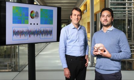
Data from the paper reveal a sharp but brief increase in neural activity– an aesthetically evokied capacity– when a stimulus pattern is shown to a mouse at about 80 milliseconds (bright orange vertical line). Notably when a stimulus is familiar, activity decreases considerably (cooler colors) after that short-term increase. Credit: Bear Lab/MIT Picower Institute
” Yet we do not yet have a clear photo of how this fundamental type of knowing is implemented within the mammalian brain,” composed Picower Professor Mark Bear and fellow authors of the new research study in the Journal of Neuroscience.
As far back as 1991 scientists discovered that when animals viewed something familiar, neurons in the cortex, or the outer layer of their brain, would be less activated than if they saw something brand-new (two of that research studys authors later on ended up being Bears coworkers at MIT, Picower Professor Earl K. Miller and Doris and Don Berkey Professor Bob Desimone).
in 2003, Bears lab occurred to observe the reverse: Mice would actually show a sharp dive in neural activity in the main visual region of the cortex when a familiar stimulus was flashed in front of the animal. This spike of activity is called a “visually evoked potential” (VEP), and Bears lab has because shown that increases in the VEPs are strong signs of VRM.
The findings in the new research study, led by previous Bear Lab postdocs Dustin Hayden and Peter Finnie, discuss how VEPs increase even amid a general decrease in neural action to familiar stimuli (as seen by Miller and Desimone), Bear stated. They also discuss more about the systems underlying VRM– the short-lived boost of a VEP might be excitation that hires inhibition, thus reducing activity overall.
New understanding
Bears lab evokes VEPs by revealing mice a black-and-white striped grating in which the stripes periodically switch their shade so that the pattern appears to reverse. Over several days as mice see this stimulus pattern, the VEPs increase, a reputable correlate of the mice becoming knowledgeable about– and less thinking about– the pattern. For 20 years Bears laboratory has actually been examining how the synapses associated with VRM modification by studying a phenomenon theyve dubbed “stimulus-selective action plasticity” (SRP).
Early studies recommended that SRP takes place among excitatory nerve cells in layer 4 of the visual cortex and particularly may need the molecular activation of their NMDA receptors.
The lab had actually seen that knocking out the receptors throughout the visual cortex prevented the increase in VEPs and for that reason SRP, however a follow-up in 2019 found that knocking them out simply in layer 4 had no effect. So, in the new research study, they decided to study VEPs, SRP, and VRM throughout the entire visual cortex, layer by layer, in search of how all of it works.
What they discovered was that a lot of the hallmarks of VRM, consisting of VEPs, occur in all layers of the cortex however that it appeared to depend on NMDA receptors on a population of excitatory neurons in layer 6, not layer 4. This is an intriguing finding, the authors stated, due to the fact that those nerve cells are well connected to the thalamus (a much deeper brain area that communicates sensory info) and to inhibitory neurons in layer 4, where they had very first measured VEPs.
They likewise measured changes in brain waves in each layer that verified a previous finding that when the stimulus pattern is new, the prevailing brain wave oscillations remain in a higher “gamma” frequency that depends on one kind of repressive neuron, however as it becomes more familiar, the oscillations shift towards a lower “beta” frequency that depends on a different inhibitory population.
A brief spike amidst a long lull
The teams rigorous and precise electrophysiology recordings of neural electrical activity in the various layers likewise revealed a prospective resolution to the contradiction between VEPs and the measures of laboratories like that of Miller and Desimone.
” What this paper reveals is that everyone is right,” Bear quipped.
How so? The brand-new data reveal that VEPs are short-term but really noticable spikes of neural electrical activity that occur amid a broader, overall lull of activity. Due to the fact that they have not had the temporal resolution to detect the quick spike, previous studies have shown just the total reduction. Bears team, on the other hand, has seen the VEPs for many years however didnt always concentrate on the surrounding lull.
The brand-new proof suggests that whats taking place is that VEP suggests the activity of the brain quickly acknowledging a familiar stimulus and then activating an inhibition of activity associated to it.
” What I think is amazing about this is that it suddenly clarifies the mechanism, due to the fact that its not that the encoding of familiarity is explained by the anxiety of excitatory synapses,” Bear said. “Rather, it appears to be accounted for by the potentiation of excitatory synapses on to nerve cells that then recruit inhibition in the cortex.”
Even as it advances that understanding of how VRM develops, the research study still leaves open questions consisting of the exact circuits included. The specific contribution of the layer 6 circuit nerve cells is not yet clear, Bear said. Therefore, the mission goes on.
Reference: “Electrophysiological signatures of visual acknowledgment memory across all layers of mouse V1” by Dustin J. Hayden, Peter S.B. Finnie, Aurore Thomazeau, Alyssa Y. Li, Samuel F. Cooke and Mark F. Bear, 15 September 2023, JNeurosci.DOI: 10.1523/ JNEUROSCI.0090-23.2023.
In addition to Hayden, Finnie, and Bear, the papers other authors are Aurore Thomazeau, Alyssa Li, and Samuel Cooke.
The National Eye Institute of the National Institutes of Health, The Picower Institute for Learning and Memory, and The JPB Foundation funded the research study.
A new study clarifies previous conflicting observations on visual acknowledgment memory (VRM), showing that increased visual stimulated potentials (VEPs) during the acknowledgment of familiar stimuli signal the brains rapid recognition procedure, eventually resulting in decreased overall neural activity.
Given that identifying what we observe as familiar or new is essential for prioritizing our attention, neuroscientists have actually devoted years to understanding why our brains excel at this task.
Throughout their research, they have actually encountered seemingly conflicting findings. Nevertheless, a recent study reveals that these difficult outcomes are truly two sides of the very same coin, leading the way for a long-sought understanding of “visual acknowledgment memory” (VRM).
VRM is the ability to rapidly acknowledge the familiar things in scenes, which can then be de-prioritized so that we can focus on the new things that might be more crucial in a given moment.