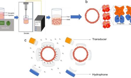
Located at the base of intestinal crypts, stem cells regenerate the numerous cell types that make up the digestive tract epithelia.During drug development, scientists require to perform appropriate nonclinical screening to make sure a drugs safety and efficacy before beginning clinical trials. A crucial aspect they must assess is a drugs pharmacokinetic properties, which explain how the compound will travel through the body to exert its activity including its absorption, metabolism, excretion, and distribution (ADME). The intestinal (GI) system is an essential system for studying a drugs ADME characteristics due to the fact that it is the main absorption website for orally administered drugs.1 As an effect, clinicians commonly observe drug-induced negative intestinal occasions within clients enrolled in scientific trials.2 Although scientists use animal and in vitro models throughout nonclinical development to examine for negative events, many GI unfavorable events are not found until human scientific trials.Difficulties with Current Nonclinical Intestinal ModelsAnimal modelsResearchers primarily count on animal designs consisting of rodents, dogs, pigs, and non-human primates for ADME and toxicity assessments due to the fact that they permit scientists to assess a drugs result in a complex system that integrates multiple organs and tissue types.3 However, researchers are typically unable to properly theorize ADME and toxicity data acquired utilizing animal models to people due to distinctions in anatomy, microbiome, physiology, and diet plan. This major drawback has actually led scientists to establish much better human in vitro designs. In vitro modelsWithin the human GI system, stem cells located in the intestinal tract crypts renew the numerous cell types that make up the epithelial layer every three to 4 days.4 Because differentiated digestive tract cells have a short lifespan, it is difficult for scientists to culture mature main epithelial cells in vitro.5 Instead, lots of use celebrated human cell lines, such as Caco-2, to study barrier stability and drug transportation across the digestive epithelia. Caco-2 cells look like fully grown enterocytes when grown as a monolayer due to their polarity, absorption abilities, and expression of some of the tight junctions, transporters, and enzymes discovered in vivo.6 However, researchers derived the Caco-2 cell line from a colon carcinoma growth and, consequently, the design shows residential or commercial properties not associated with healthy intestinal tract epithelia. Furthermore, Caco-2 monocultures prevent researchers from evaluating the drugs impact on other digestive cell types, such as goblet cells or enteroendocrine cells.When scientists culture intestinal stem cells separated from native intestinal tract crypts in the existence of extracellular matrix proteins and growth elements, the cells differentiate and form organoids, where their internal cavity represents the intestinal lumen.7 These three-dimensional in vitro models reproduce the functionality and structure of intestinal tracts in vivo much better than Caco-2 monocultures and enable scientists to evaluate a drugs effect on several cell types. However, the organoids apical surface deals with the internal cavity, which forces scientists to jeopardize the epithelial layer when injecting drugs into its lumen. This avoids them from evaluating barrier stability and from carrying out high-throughput analyses. Scientists can circumvent these problems by reversing the organoids polarity, such that the apical surface deals with towards the culture medium. Nonetheless, scientists are unable to quickly access the apical and basolateral surfaces concurrently when using organoids in either confirmation. Generating an Advanced In Vitro Intestinal Model Despite making use of these available designs throughout nonclinical screening, scientists are typically unable to spot drug-induced gastrointestinal toxicity or accurately examine its absorption up until they carry out medical trials, likely since the designs are not physiologically pertinent, predictive of the human response, or favorable to high-throughput screening. Appropriately, scientists are still in search of much better model systems to prevent late-stage medical failures.The RepliGut ® Systems from Altis Biosystems are a group of in vitro, nonclinical models that replicate the human intestinal epithelia. Like organoids, these systems utilize human donor-extracted digestive tract stem cells, which are seeded onto scaffold-coated, semi-permeable membranes to form monolayers. Upon distinction, researchers acquire the significant digestive cell family trees at the very same proportion found within the body. Because of the systems speculative set-up, scientists can easily access both the basolateral and apical surface areas of the digestive epithelium, which is a major limitation of 3D organoid cultures.8 Additionally, Altis Biosystems has extracted stem cells from several regions of the GI tract and from several donors. This allows researchers to examine drug toxicity and absorption, along with the integrity of the epithelial cell barrier, across various areas and donors. These physiologically-relevant in vitro designs are basic to use, suitable with high-throughput analysis, and save scientists money and time, thus, accelerating nonclinical research.ReferencesAzman M, et al. Intestinal tract absorption study: Challenges and absorption improvement strategies in improving oral drug delivery. Pharm Basel Switz. 2022; 15( 8 ):975. Peters MF, et al.. Establishing in vitro assays to change gastrointestinal security assessment: Potential for microphysiological systems. Lab Chip. 2020; 20( 7 ):1177 -1190. Henze LJ, et al. The pig as a preclinical design for anticipating oral bioavailability and in vivo performance of pharmaceutical oral dosage forms: a PEARRL review. J Pharm Pharmacol. 2019; 71( 4 ):581 -602. Rees WD, et al. Regenerative intestinal tract stem cells induced by chronic and acute injury: The saving grace of the epithelium? Front Cell Dev Biol. 2020; 8. Wang Y, et al. Self-renewing monolayer of main colonic or rectal epithelial cells. Cell Mol Gastroenterol Hepatol. 2017; 4( 1 ):165 -182. e7.van Breemen RB, Li Y. Caco-2 cell permeability assays to determine drug absorption. Expert Opin Drug Metab Toxicol. 2005; 1( 2 ):175 -185. Sato T, et al. Single Lgr5 stem cells construct crypt-villus structures in vitro without a mesenchymal niche. Nature. 2009; 459( 7244 ):262 -265. Pike CM, et al. Characterization and optimization of variability in a human colonic epithelium culture design. Preprint. bioRxiv. Released online September 22, 2023:2023.09.22.559007..
Located at the base of intestinal crypts, stem cells regrow the various cell types that make up the digestive epithelia.During drug development, researchers require to perform sufficient nonclinical screening to make sure a drugs security and effectiveness before beginning medical trials. In vitro modelsWithin the human GI tract, stem cells located in the digestive tract crypts replenish the different cell types that make up the epithelial layer every three to 4 days.4 Because differentiated intestinal tract cells have a brief life expectancy, it is difficult for researchers to culture fully grown primary epithelial cells in vitro.5 Instead, many utilize immortalized human cell lines, such as Caco-2, to study barrier integrity and drug transportation across the intestinal epithelia. Caco-2 cells look like mature enterocytes when grown as a monolayer due to their polarity, absorption abilities, and expression of some of the tight junctions, transporters, and enzymes discovered in vivo.6 However, researchers derived the Caco-2 cell line from a colon cancer growth and, subsequently, the design shows properties not associated with healthy digestive epithelia. Additionally, Caco-2 monocultures prevent researchers from assessing the drugs impact on other digestive tract cell types, such as goblet cells or enteroendocrine cells.When scientists culture intestinal stem cells separated from native digestive tract crypts in the existence of extracellular matrix proteins and growth factors, the cells differentiate and form organoids, where their internal cavity represents the digestive tract lumen.7 These three-dimensional in vitro models replicate the performance and structure of intestinal tracts in vivo much better than Caco-2 monocultures and make it possible for scientists to check a drugs result on several cell types. Due to the fact that of the systems speculative set-up, scientists can easily access both the apical and basolateral surfaces of the intestinal epithelium, which is a major limitation of 3D organoid cultures.8 Additionally, Altis Biosystems has actually drawn out stem cells from a number of regions of the GI tract and from multiple donors.