

MIT researchers have created a wearable ultrasound patch that can non-invasively image internal organs, initially focusing on bladder health. This device, which does not require an ultrasound operator or gel, has the potential to revolutionize the monitoring of various organ functions and disease detection.
The wearable device, designed to monitor bladder and kidney health, could be adapted for earlier diagnosis of cancers deep within the body.
MIT researchers have designed a wearable ultrasound monitor, in the form of a patch, that can image organs within the body without the need for an ultrasound operator or application of gel.
In a new study, the researchers showed that their patch can accurately image the bladder and determine how full it is. This could help patients with bladder or kidney disorders more easily track whether these organs are functioning properly, the researchers say.
This approach could also be adapted to monitor other organs within the body by changing the location of the ultrasound array and tuning the frequency of the signal. Such devices could potentially enable earlier detection of cancers that form deep within the body, such as ovarian cancer.
“This technology is versatile and can be used not only on the bladder but any deep tissue of the body. It’s a novel platform that can do identification and characterization of many of the diseases that we carry in our body,” says Canan Dagdeviren, an associate professor in MIT’s Media Lab and the senior author of the study.
Lin Zhang, an MIT research scientist; Colin Marcus, an MIT graduate student in electrical engineering and computer science; and Dabin Lin, a professor at Xi’an Technological University, are the lead authors of a paper describing the work, which was published recently in Nature Electronics.
Wearable Monitoring
Dagdeviren’s lab, which specializes in designing flexible, wearable electronic devices, recently developed an ultrasound monitor that can be incorporated into a bra and used to screen for breast cancer. In the new study, the team used a similar approach to develop a wearable patch that can adhere to the skin and take ultrasound images of organs located within the body.
For their first demonstration, the researchers decided to focus on the bladder, partly inspired by Dagdeviren’s younger brother, who was diagnosed with kidney cancer a few years ago. After having one of his kidneys surgically removed, he had difficulty fully emptying his bladder. Dagdeviren wondered if an ultrasound monitor that reveals how full the bladder is might help patients similar to her brother, or people with other types of bladder or kidney problems.
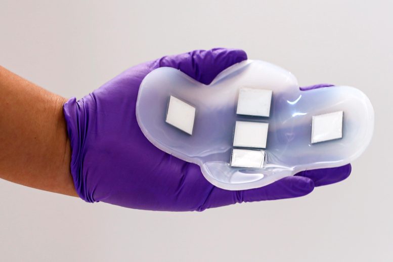
A wearable ultrasound monitor can image the bladder and determine how full it is. The MIT-developed device could help patients with bladder or kidney disorders more easily track whether these organs are functioning properly. Credit: Courtesy of the researchers
“Millions of people are suffering from bladder dysfunction and related diseases, and not surprisingly, bladder volume monitoring is an effective way to assess your kidney health and wellness,” she says.
Currently, the only way to measure bladder volume is using a traditional, bulky ultrasound probe, which requires going to a medical facility. Dagdeviren and her colleagues wanted to develop a wearable alternative that patients could use at home.
To achieve that, they created a flexible patch made of silicone rubber, embedded with five ultrasound arrays made from a new piezoelectric material that the researchers developed for this device. The arrays are positioned in the shape of a cross, which allows the patch to image the entire bladder, which is about 12 by 8 centimeters when full.
The polymer that makes up the patch is naturally sticky and adheres gently to the skin, making it easy to attach and detach. Once placed on the skin, underwear or leggings can help to hold it in place.
Clinical Study and Results
In a study performed with collaborators at the Center for Ultrasound Research and Translation and Department of Radiology at Massachusetts General Hospital, the researchers showed that the new patch could capture images comparable to those taken with a traditional ultrasound probe, and these images could be used to track changes in bladder volume.
For the study, the researchers recruited 20 patients with a range of body mass indexes. Subjects were first imaged with a full bladder, then with a partially empty bladder, and then with a completely empty bladder. The images obtained from the new patch were similar in quality to those taken with traditional ultrasound, and the ultrasound arrays worked on all subjects regardless of their body mass index.
Using this patch, no ultrasound gel is needed, and no pressure needs to be applied, as with a regular ultrasound probe, because the field of view is large enough to encompass the entire bladder.
Future Developments and Goals
To see the images, the researchers connected their ultrasound arrays to the same kind of ultrasound machine used in medical imaging centers. However, the MIT team is now working on a portable device, about the size of a smartphone, that could be used to view the images.
“In this work, we have further developed a path toward clinical translation of conformable ultrasonic biosensors that yield valuable information about vital physiologic parameters. Our group hopes to build on this and develop a suite of devices that will ultimately bridge the information gap between clinicians and patients,” says Anthony E. Samir, director of the MGH Center for Ultrasound Research and Translation and Associate Chair of Imaging Sciences at MGH Radiology, who is also an author of the study.
The MIT team also hopes to develop ultrasound devices that could be used to image other organs within the body, such as the pancreas, liver, or ovaries. Based on the location and depth of each organ, the researchers need to alter the frequency of the ultrasound signal, which requires designing new piezoelectric materials. For some of these organs, located deep within the body, the device may work better as an implant rather than a wearable patch.
“For whatever organ that we need to visualize, we go back to the first step, select the right materials, come up with the right device design, and then fabricate everything accordingly,” before testing the device and performing clinical trials, Dagdeviren says.
“This work could develop into a central area of focus in ultrasound research, motivate a new approach to future medical device designs, and lay the groundwork for many more fruitful collaborations between materials scientists, electrical engineers, and biomedical researchers,” says Anantha Chandrakasan, dean of MIT’s School of Engineering, the Vannevar Bush Professor of Electrical Engineering and Computer Science, and an author of the paper.
Reference: “A conformable phased-array ultrasound patch for bladder volume monitoring” by Lin Zhang, Colin Marcus, Dabin Lin, David Mejorado, Scott Joseph Schoen Jr, Theodore T. Pierce, Viksit Kumar, Sara V. Fernandez, David Hunt, Qian Li, Ikra Iftekhar Shuvo, David Sadat, Wenya Du, Hannah Edenbaum, Li Jin, Weiguo Liu, Yonina C. Eldar, Fei Li, Anantha P. Chandrakasan, Anthony E. Samir and Canan Dagdeviren, 16 November 2023, Nature Electronics.
DOI: 10.1038/s41928-023-01068-x
The research was funded by a National Science Foundation CAREER award, a 3M Non-Tenured Faculty Award, the Sagol Weizmann-MIT Bridge Program, Texas Instruments Inc., the MIT Media Lab Consortium, a National Science Foundation Graduate Research Fellowship, and an ARRS Scholar Award.