
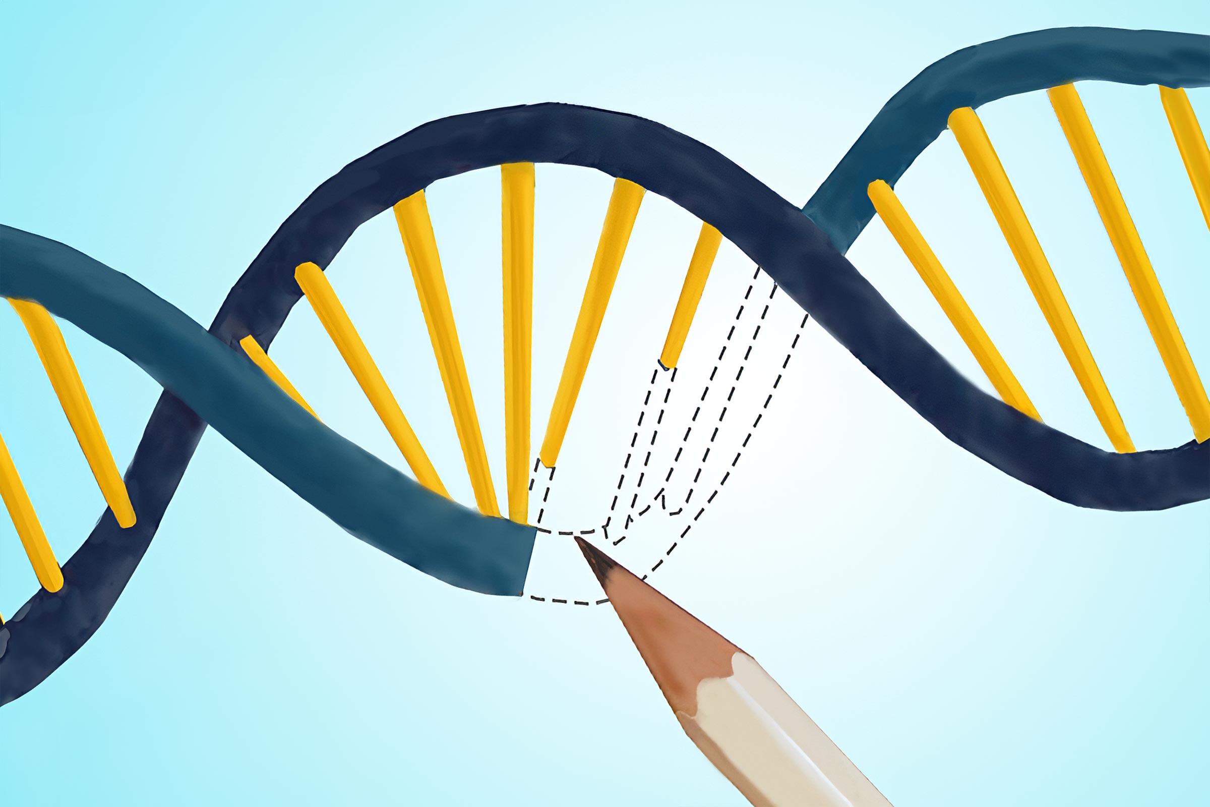
Although the specificity of CRISPR-based gene-editing is highly accurate and versatile, the efficiency of installing those edits has been low. In this paper, the Adamson lab describes a more efficient prime editor. Credit: Caitlin Sedwick for Princeton University
Princeton scientists make a major improvement to a CRISPR-based gene-editing tool called “prime editing.”
Through years of engineering gene-editing systems, researchers have developed a suite of tools that enable the modification of genomes in living cells, akin to ‘genome surgery’. These tools, including ones based on a natural system known as CRISPR/Cas9, offer enormous potential for addressing unmet clinical needs, underscored by the recent FDA approval of the first CRISPR/Cas9-based therapy.
A relatively new approach called “prime editing” enables gene-editing with exceptional accuracy and high versatility, but has a critical tradeoff: variable and often low efficiency of edit installation. In other words, while prime edits can be made with high precision and few unwanted byproducts, the approach also often fails to make those edits at reasonable frequencies.
Enhanced Prime Editing Techniques
In a paper that appeared in print in the journal Nature on April 18th, 2024, Princeton scientists Jun Yan and Britt Adamson, along with several colleagues, describe a more efficient prime editor.

Authors (l-r) Brittany Adamson, Assistant Professor of Molecular Biology and the Lewis-Sigler Institute for Integrative Genomics; and Jun Yan, Adamson lab graduate student and first author. Credit: Photo of Britt Adamson by Denise Applewhite, Princeton University. Photo of Jun Yan by the author.
Prime editing systems minimally consist of two components: a modified version of the protein element of CRISPR/Cas9 and a ribonucleic acid (RNA) molecule called a pegRNA. These components work together in several coordinated steps: First, the pegRNA binds the protein and guides the resulting complex to a desired location in the genome. There, the protein nicks the DNA and, using a template sequence encoded on the pegRNA, “reverse transcribes” an edit into the genome nearby. In this way, prime editors “write” exact sequences into targeted DNA.
“Prime editing is such an incredibly powerful genome editing tool because it gives us more control over exactly how genomic sequences are changed,” Adamson said.
Experimental Insights and Innovations
At the outset of their study, Adamson and Yan, a graduate student in Adamson’s research group and the Department of Molecular Biology, reasoned that unknown cellular processes may aid or hinder prime editing. To identify such processes, Yan laid out a conceptually simple plan: First, he would engineer a cell line that would emit green fluorescence when certain prime edits were installed. Then, he would systematically block expression of proteins normally expressed within those cells and measure editing-induced fluorescence to determine which of those proteins impact prime editing. By executing this plan, the team identified 36 cellular determinants of prime editing, only one of which—the small RNA-binding protein La—promoted editing.
“Although promoting prime editing is obviously not a normal function of the La protein, our experiments showed that it can strongly facilitate the process,” Yan said.
Within cells, La is known to bind specific sequences often found at the ends of nascent small RNA molecules and it protects those RNAs from degradation. The Princeton team recognized right away that the pegRNAs deployed in Yan’s first experiments likely contained those exact sequences, called polyuridine tracts, as they are a typical but often overlooked byproduct of pegRNA expression in cells. Subsequent experiments suggested that such pegRNAs inadvertently harness La’s end-binding activity for protection and to promote prime editing.
Development of the PE7 Protein
Motivated by their results, the team asked if fusing the part of La that binds polyuridine tracts to a standard prime editing protein could boost prime editing efficiencies. They were thrilled to find that the resulting protein, which they call PE7, substantially enhanced intended prime editing efficiencies across conditions and, when using some prime editing systems, left the frequencies of unwanted byproducts very low. Their results quickly drew the attention of colleagues interested in using prime editing in primary human cells, including Daniel Bauer at Boston Children’s Hospital and Harvard Medical School and Alexander Marson at the University of California, San Francisco. Together with scientists from these labs, the team of researchers went on to demonstrate that PE7 can also enhance prime editing efficiencies in therapeutically relevant cell types, offering expanded promise for future clinical applications.
“This work is a beautiful example of how deeply probing the inner workings of cells can lead to unexpected insights that may yield near-term biomedical impact,” Bauer noted.
Reference: “Improving prime editing with an endogenous small RNA-binding protein” by Jun Yan, Paul Oyler-Castrillo, Purnima Ravisankar, Carl C. Ward, Sébastien Levesque, Yangwode Jing, Danny Simpson, Anqi Zhao, Hui Li, Weihao Yan, Laine Goudy, Ralf Schmidt, Sabrina C. Solley, Luke A. Gilbert, Michelle M. Chan, Daniel E. Bauer, Alexander Marson, Lance R. Parsons and Britt Adamson, 3 April 2024, Nature.
DOI: 10.1038/s41586-024-07259-6
Funding: Funding for this work was provided by the National Institutes of Health (NIH) (R35GM138167, RM1HG009490, T32HG003284, DP2CA239597, UM1HG012660 [Princeton QCB training grant; NHGRI], and [T32GM007388 Princeton MOL training grant; NIGMS]); the Searle Scholars Program; the Princeton Catalysis Initiative; CHDI Foundation; Princeton University; the Parker Institute for Cancer Immunotherapy (PICI); the Lloyd J. Old STAR award from the Cancer Research Institute (CRI); the Simons Foundation; the CRISPR Cures for Cancer Initiative; the Arc Institute; CRUK/NIH (OT2CA278665 and CGCATF-2021/100006); Pew-Stewart Scholars for Cancer Research award; the Doris Duke Foundation; the St Jude Children’s Research Hospital Collaborative Research Consortium; NHLBI (R01HL150669); the Fred Hutch Cooperative Center of Excellence in Hematology (U54 DK106829); the China Scholarship Council (CSC), based on the April 2015 Memorandum of Understanding between the CSC and Princeton University; the NCI (K00CA245718); and the Princeton University Flow Cytometry Resource Facility (NCI-CCSG P30CA072720-5921).
Grant numbers: R35GM138167, RM1HG009490, T32HG003284, DP2CA239597, UM1HG012660, T32GM007388, OT2CA278665, CGCATF-2021/100006, U54 DK106829, K00CA245718, NCI-CCSG P30CA072720-5921
Funders: National Institutes of Health (NIH), Searle Scholars Program, Princeton Catalysis Initiative, CHDI Foundation; Princeton University, Parker Institute for Cancer Immunotherapy (PICI), Cancer Research Institute (CRI), Simons Foundation, CRISPR Cures for Cancer Initiative, Arc Institute, CRUK/NIH, Pew-Stewart, Doris Duke Foundation, St Jude Children’s Research Hospital Collaborative Research Consortium, NHLBI, Fred Hutch Cooperative Center of Excellence in Hematology, China Scholarship Council (CSC), NCI.