” The cell wall that lines the ventricles wears out in time, like a balloon thats consistently exploded and deflated,” stated Weickenmeier. “And the stresses arent consistent– theyre defined by the geometry of the ventricle, so we can anticipate where these failures will occur.”
The model provides an easy, physics-based explanation for the areas of these lesions, revealing that mechanical loads “need to be a significant contributor to the onset of illness,” stated Weickenmeier.
The teams research study, published just recently in Scientific Reports, used 2D imaging showing a cross-section of the brain, but Weickenmeiers team has since expanded its research study to a complete 3D design of the brain. Next, Weickenmeier hopes to utilize advanced MRI technologies developed at Stevens to study the motion of the ventricle wall directly.
Johannes Weickenmeier in the Experimental and Computational Soft Matter Biomechanics Lab at Stevens Institute of Technology Credit: Stevens Institute of Technology.
In the long term, the teams findings may allow the advancement of brand-new treatments for sores. Normally, pharmaceutical treatments struggle to reach and cross the blood-brain barrier affected areas, but the brand-new research recommends that it may be possible to channel drugs to sores directly through leaks in the ventricular wall.
The more comprehensive takeaway from the groups research study, discussed Weickenmeier, is that the brains aging procedure is moderated by physical processes, consisting of the pressure of distributing blood and CSF. That underscores the requirement for healthy behaviors– such as getting adequate exercise and avoiding harmful compounds– that can decrease those strains on the brain.
Reference: “Peak ependymal cell stretch overlaps with the start locations of periventricular white matter sores” by Valery L. Visser, Henry Rusinek and Johannes Weickenmeier, 9 November 2021, Scientific Reports.DOI: 10.1038/ s41598-021-00610-1.
Generally, pharmaceutical treatments battle to reach and cross the blood-brain barrier affected locations, however the brand-new research suggests that it may be possible to transport drugs to sores directly through leakages in the ventricular wall.
Due to the fact that they reveal up as intense white spots on MRI scans– are improperly understood, these sores– known as periventricular and deep white matter hyperintensities. However they are not unusual: the majority of people have some by the time they reach their 60s, and modifications only increase with age. The more lesions that build up and the quicker they grow, the more susceptible we become to cognitive problems ranging from memory issues to motor disorders.
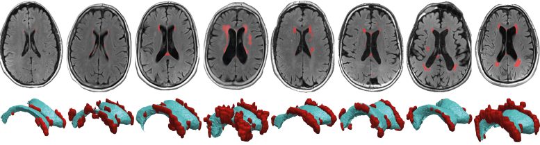
Using MRI scans from 8 healthy topics, Weickenmeier worked with Valery Visser, now a doctorate student at the University of Zurich, and Henry Rusinek, a radiologist at NYU Grossman School of Medicine, to develop a personalized computer system model of each subjects brain. The group mapped the pressure put on ventricular walls, the linings of fluid-filled chambers deep in the brain, as waves of pressure pulse through the topics cerebral spine fluid, or CSF. They found that hyperintensities tend to happen near locations that should extend more to accommodate pressure changes of the distributing CSF because, as such areas wear thin, CSF can leakage into the brain and trigger sores.
When the walls of the CSF-filled ventricle (black) wear thin, CSF leakages into the brain tissue (grey) and develops sores. As our brains age, small lesions begin to pop up in the packages of white matter that bring messages in between our neurons. Now, researchers at Stevens Institute of Technology and colleagues not just offer a description for the area of these lesions however also how they establish in the very first place.
They discovered that hyperintensities tend to occur near locations that must extend more to accommodate pressure modifications of the flowing CSF due to the fact that, as such areas use thin, CSF can leakage into the brain and trigger lesions.
Lesions (red) happen near areas that need to stretch more to accommodate pressure modifications of the flowing cerebrospinal fluid. When the walls of the CSF-filled ventricle (black) wear thin, CSF leakages into the brain tissue (grey) and creates lesions. Credit: Stevens Institute of Technology
Scientists at Stevens Institute of Technology show that strain on ventricular walls describes where sores establish in the aging brain.
As our brains age, little lesions begin to turn up in the packages of white matter that bring messages between our neurons. The sores can damage this white matter and lead to cognitive deficits. Now, scientists at Stevens Institute of Technology and coworkers not only supply a description for the area of these lesions however likewise how they establish in the very first location.
The work, led by Johannes Weickenmeier, an assistant teacher of mechanical engineering at Stevens, highlights the significance of viewing the brain as more than neural circuitry that underpins how ideas are formed, and memories produced. Its also a physical object thats susceptible to glitches and mechanical failures. “The brain is susceptible to tear and use in vulnerable areas,” Weickenmeier said. “Especially in an aging brain, we need to look at its biomechanical homes to much better comprehend how things can begin to fail.”