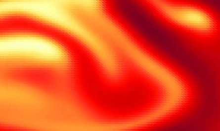
On the surface area, viral glycoproteins (red) are incorporated into a membrane (transparent). The membrane encloses various viral tegument proteins (gray) and assimilated host proteins (pink). The viral tegument protein UL32 is highlighted in yellow.
Herpes viruses are treacherous: as soon as they infect you, they remain with you for life. Nearly every adult unwittingly hosts at least one of the nine unique human herpes viruses.
Herpes infections are so effective due to the fact that of their amazing adaptation to humans and their strategies to evade our immune system. Secret to their camouflage are proteins that deceive the contaminated cell into thinking its not under hazard. It is understood, for example, that every herpes infection has an effective proteome, i.e. a great deal of these proteins, which, highly adapted to the host, allows it to reproduce effectively right away after infection.
These recently formed infections– likewise called virions– contain various viral proteins as well as host proteins. A layer of numerous other proteins called tegument is formed around this capsid.
Particles come into play in the reactivation of the infection
The particles are important in enabling the virus to replicate again and spread out systemically in the body after reactivation set off by whatever suggests. They are therefore main to the outbreak of illness– after an extended period of dormancy (latency).
Little is understood about the internal organization of these particles, especially the protein-protein interactions within the tegument. Researchers from the Leibniz-Forschungsinstitut für Molekulare Pharmakologie (FMP) and the Charité– Universitätsmedizin Berlin have therefore taken a more detailed look at the particles, specifically in human cytomegalovirus (HCMV).
HCMV happens particularly often in the population and can be actually harmful, especially for transplant receivers and unborn children who end up being contaminated through the mom. Regardless of extensive research, there is currently no well-tolerated antiviral treatment that might efficiently control or even remove the virus. There is also no vaccination versus this type of virus.
Map programs which proteins interact with each other
In the present work, the group led by Fan Liu (FMP) and Lüder Wiebusch (Charité) has for the very first time produced a detailed map of the spatial interactions in between viral and host cell proteins within HCMV particles. Among other things, this revealed that specific host cell proteins are hired by viral proteins and play a function in viral replication. For example, a viral protein called UL32 hires a cellular protein (protein phosphatase, PP1) into the particle to prevent binding of other, undesirable, host cell proteins.
” HCMV itself does not have any phosphatases like PP1, so you can see that the infection makes the most of particular host cell proteins to duplicate efficiently,” states FMP virologist Boris Bogdanow, discussing a crucial technique for how HCMV techniques its host.
To study the interactions between the various proteins in intact HCMV particles layer by layer, the researchers used a strategy called cross-linking mass spectrometry. “This approach also enables us to reason about the identity of the proteins,” highlighted Fan Liu, a professional in mass spectrometry at the FMP. “But what is special and special about cross-linking is that we can see which proteins engage with each other and where.”
Never ever before has this innovative innovation been used to map the spatial company of interactions within herpesviral particles. With the information thus obtained, a computer system design of the HCMV particle was subsequently created at FU Berlin by Mohsen Sadeghi. The virtual model allows the simulation of each protein within the particle and imagines the biophysical processes in a vibrant method.
” The identified protein-protein interaction is crucial to better understand the complicated life process of HCMV,” Boris Bogdanow classifies the results. “And this, in turn, is very important for discovering candidate anti-viral drugs versus HCMV.”
Referral: “Spatially solved protein map of undamaged human cytomegalovirus virions” by Boris Bogdanow, Iris Gruska, Lars Mühlberg, Jonas Protze, Svea Hohensee, Barbara Vetter, Jens B. Bosse, Martin Lehmann, Mohsen Sadeghi, Lüder Wiebusch and Fan Liu, 7 August 2023, Nature Microbiology.DOI: 10.1038/ s41564-023-01433-8.
The membrane encloses different viral tegument proteins (gray) and absorbed host proteins (pink). These recently formed infections– also called virions– consist of various viral proteins as well as host proteins. In the existing work, the team led by Fan Liu (FMP) and Lüder Wiebusch (Charité) has for the first time produced a comprehensive map of the spatial interactions in between viral and host cell proteins within HCMV particles. Among other things, this revealed that certain host cell proteins are hired by viral proteins and play a function in viral replication. A viral protein called UL32 recruits a cellular protein (protein phosphatase, PP1) into the particle to avoid binding of other, undesirable, host cell proteins.